Biology Reference
In-Depth Information
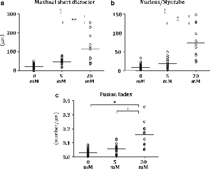
Fig. 29.1
The effect of taurine treatment on differentiation of C2C12 myoblast to myotube. C2C12
cells were cultured with 0, 5, or 20 mM taurine in the differentiation medium for 7 days. (
a
) The length
of maximum short diameter in myotube, (
b
) the number of nucleus per myotube, and (
c
) fusion index
was calculated by the number of nucleus per the examined myotube length. Data are shown as the
median and value plots. *
p
< 0.05, **
p
< 0.01, ‡
p
< 0.0001 by one-way ANOVA analysis
Likewise, the number of nucleus in the differentiated myotube (Fig.
29.1b
) and
fusion index (Fig.
29.1c
) were also significantly increased by 20 mM taurine treat-
ment compared to that in the control and 5 mM taurine treatment.
29.3.2
The Effects of Inhibitors of Taurine Transport and Ca
+2
Signaling on the Taurine-Enhanced C2C12 Differentiation
Figure
29.2
shows the fluorescence images of C2C12 myotube treated with nife-
dipine or transfected with
taut
siRNA. During the differentiation period, the cells
in both conditions were exposed to 20 mM taurine. The enhanced effect of taurine
on the differentiation to myotube evaluated by FITC-positive MHC protein expres-
sion was cancelled by the treatment of nifedipine (Fig.
29.2a
). The expression of
MCIP-1 mRNA in myotube was significantly increased by nifedipine treatment
compared to that in undifferentiated myoblast, and the expression level in myotube
Search WWH ::

Custom Search