Biology Reference
In-Depth Information
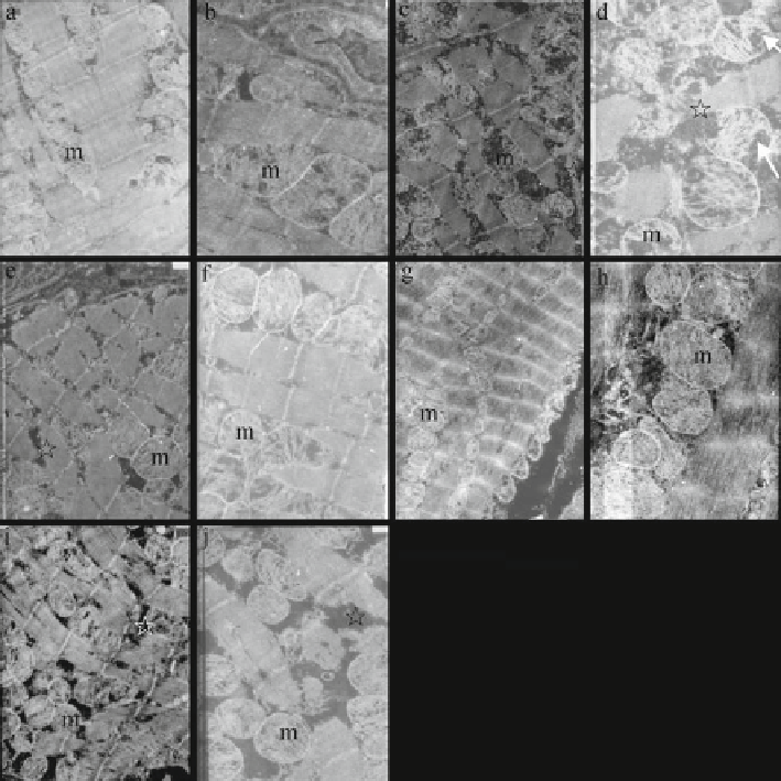
Fig. 21.5
The ultrastructural changes of cardiomyocytes from the left ventricular at the level of the
near apex. (
a
,
b
) Group C showed fine dispersed nuclear chromatin, slightly contracted myofibrils,
well-arranged Z disk, mitochondria with tightly packed cristae, and an intact sarcolemma. (
c
,
d
)
Model group showed disarrayed myofibrils with obvious fragmentation and dissolution; curved
Z-line; swollen mitochondria with obvious amorphous matrix densities, some cristae vanishing, and
disruption of mitochondrial membranes; ruptured sarcolemma; and edema of sarcoplasm. (
e
,
f
)
Group PI showed a lesser extent injury than the model group. (
g
,
h
) Group PII showed results simi-
lar to those described in control group. (
i
,
j
) Group P III showed the structure of mitochondria was
preserved by taurine administration compared with model group. Magnification (as indicated): (
a
,
c
,
e
,
g
,
i
) ×8,000; (
b
,
d
,
f
,
h
,
j
) ×15,000. Scale bars = 1.25 mm (×8,000) and 0.67 mm (×15,000).
M
mitochondrion,
arrows
mitochondria breakdown,
asterisk
myo fi lament breakdown
described in control group. The cardiomyocytes in group PIII (Fig.
21.5i, j
) also
showed obvious edema of sarcoplasm, disarrayed mitochondria that are slightly
swollen, and myofibrils fragmentation and dissolution compared with control
group. However the structure of mitochondria was preserved by taurine compared
with model group. The results indicated 200 mg/kg taurine had better protective
effect.
Search WWH ::

Custom Search