Biomedical Engineering Reference
In-Depth Information
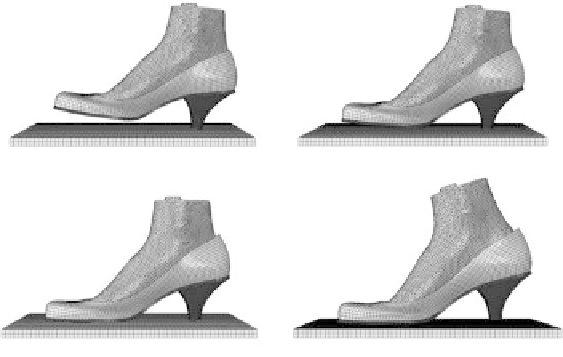
Initial contact
Heel strike
Midstance
Push off
FIgure 2.11
Finite element-predicted walking shod in high-heeled shoes.
pressure, internal stress, and movement of different foot regions and bony structures in correspond-
ing loading conditions are compared in the following subsections.
2.3.3.1 Interfacial Foot Pressure during donned and Shod walking Conditions
The FE-predicted plantar pressure distribution patterns in general showed good agreement with the
experimental measurements, while the predicted peak pressures at heel strike (0.47 MPa) and push
off (0.63 MPa) phases were 14% and 26% larger than the measured values. During heel strike, peak
pressure was located at the center of the heel. The pressurized area was concentrated at the lateral
midfoot, ball of the foot, and toes during midstance. Localized peak pressure was found underneath
the second and third metatarsal heads and at the center of the heel but there was a large area having
no contact at the forefoot region during midstance. At push off, the heel and lateral midfoot were
unloaded and the peak pressure at the forefoot intensified. The FE-predicted interfacial pressures,
including plantar and dorsal pressure, during donned and shod walking conditions are tabulated
in Table 2.1. After donning, slight contact pressure was found at the heel and toe regions, while
noticeable contact pressure concentrated at the dorsum of the first, fourth, and fifth toes. During
table 2.1
Finite element-Predicted Plantar and dorsal Foot Contact Pressure (mPa) for Simulated
high-heeled Shoe donning and walking Conditions
donned
heel strike
midstance
Push off
Foot regions
Plantar
dorsal
Plantar
dorsal
Plantar
dorsal
Plantar
dorsal
Toes
1
st
0.03
0.07(M)
0.10(S)
0.07
0.10(M)
0.10(S)
0.15
0.18(M)
0.10(S)
0.36
0.42(M)
0.11(S)
4
th
-
0.10
0.02
0.07
0.06
0.14
0.17
0.12
5
th
-
0.13
-
0.11
0.10
0.31
0.36
0.32
Forefoot
-
0.04
-
0.07
0.23
0.06
0.63
0.11
Midfoot
0.04
-
0.10
0.05
0.08
0.06
-
-
Rear foot
-
0.03
0.47
0.06
0.22
0.03
-
-
Note:
M means medial side surface of the dorsum of the first toe as shown in Figure 2.12; S means superior side surface
of the dorsum of the first toe as shown in Figure 2.12.


Search WWH ::

Custom Search