Biomedical Engineering Reference
In-Depth Information
22.4.2 a
nalySiS
of
tmd m
odelS
and
a
pplicationS
In the base model with normal TMJ, the maximum contact stresses were located between the ante-
rior condyle and the lower surface of the intermediate zone of the disc and between the upper sur-
face of the intermediate zone of the disc and the posterior articular eminence (i.e. anterior temporal
bone), respectively. Therefore the maximum stresses occurred at the intermediate zone of the disc,
and the anterior of the condyle and temporal bone, accordant with the physiological function and
related biomechanical research (del Palomar and Doblare 2006c; Koolstra and van Eijden 2005;
Oberg et al. 1971).
22.4.2.1 relaxation of the discal attachment
Abnormal positioning of the pathologic disc with respect to the condyle and articular fossa-eminence
was caused by relaxation of disc attachments, which altered the biomechanical environment of the
pathologic side of the TMJ, but scarcely affected the asymptomatic side. Thus, the results of the
pathologic TMJs were analyzed.
The interaction between the lateral intermediate zone of the disc and the lateral side of the con-
dyle and the articular fossa-eminence was evident in the model with relaxation of the anterior disc
attachments. Therefore, the maximum stresses of the pathologic disc were located at the lateral
intermediate zone. The stress level in the pathologic disc was significantly greater than the identical
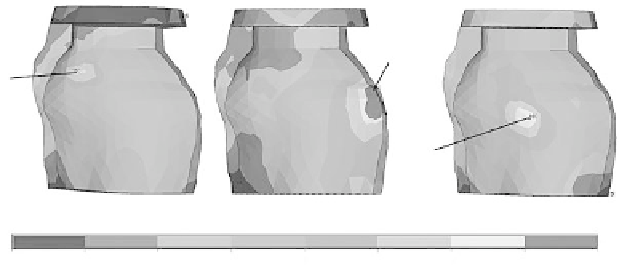
disc in the base model, as shown in Figure 22.7. The maximum compressive stress of the patho-
logic disc was about five times greater than the identical disc in the base model, and the maximum
tensile and shear stresses of the pathologic disc were about triple the magnitude of the base model.
Thinning or perforation of the lateral intermediate zone of the disc with the relaxed anterior attach-
ment may result from long-term effects of occlusion and mandibular movement. According to the
contact status, the maximum stresses of the condyle and the temporal bone were located at the
lateral region. However, the maximum stresses of the anterior of the condyle and the temporal bone
were much lower than those in the base model.
In the model with relaxation of the bilaminar zones (posterior disc attachments), the maximum
contact stress, located at the intermediate zone of the disc and posterior articular eminence, was
much greater than that of the base model. Contrarily, the interaction between the disc and the con-
dyle was weaker than that of the base model. The maximum stresses of the pathologic disc were
located at the intermediate zone. The stress level in the pathologic disc was slightly greater than the
identical disc in the base model, as shown in Figure 22.7. Based on the contact status, the maximum
stresses of the condyle and the temporal bone were located at the anterior region. The maximum
Max:
0.522MPa
Max:
0.239MPa
Max:
0.315MPa
(a)
(b)
(c)
0
.005
.05
.2
.5
.100E
-
02
.01
.1
.3
FIgure 22.7
(See color insert.)
The von Mises stress distributions of the normal disc and the discs with
relaxed attachment (the maximum stresses were listed; units: MPa): (a) normal disc, (b) disc with relaxation
of anterior attachments, and (c) disc with relaxation of bilaminar zones.
Search WWH ::

Custom Search