Biomedical Engineering Reference
In-Depth Information
in head injury. In 2003, King et al. presented a review of brain injury mechanisms with a distinction
between the effects of linear and angular acceleration on the mechanisms of concussion. An intrigu-
ing example from nature is the case of woodpeckers, who drum tree trunks at a speed of 6-7 m/s
with a linear acceleration of approximately 1000 g and an angular acceleration of 297 krad/s with
no head injuries (May et al., 1979; Wang, Cheung et al., 2011). Indeed, the woodpecker drums about
10-20 bouts continuously, and every bout takes about 50 milliseconds. It drums about 12,000 times
per day on average (Spring, 1965). Woodpeckers perform rhythmic drumming with their beaks on
surfaces such as dead tree limbs to catch and feed themselves with worms, or to attract mates and
announce their territorial boundaries (Spring, 1965). The kinematics of the woodpecker's head has
been collected in previous studies (Wang et al., 2011).
19.2 develoPment oF a FInIte element model oF
human and woodPeCker headS
Due to the fact that in real-world collisions, head injury occurs from a combination of translational
and rotational acceleration, neither of which needs to be extremely high for the current measure-
ment devices, sophisticated computer models of the head can provide useful information in the
investigation of human injury due to impact such as intracranial response under real world head
impact conditions. It was necessary to understand the head anatomy for constructing its geometrical
model.
19.2.1 H
uman
and
W
oodpecker
H
ead
a
natomy
The human head consists of different tissue layers such as the scalp, skull bone, and dural, arach-
noidal, and pial membranes, as well as cerebrospinal fluid (CSF), that cover the brain (Figure 19.1).
The skull bone can be viewed as a three-layered sandwich structure with an inner and outer table of
cortical bone and spongy bone sandwiched between them. The brain, with its covering membranes
and CSF, is connected to the spinal cord through the foramen magnum. The inferior part of the
skull base is attached to the neck by articulation through occipital condyles, ligaments, and muscles.
The woodpecker's brain is tightly packed by relatively dense yet spongy bone, especially evident
at the occiput, in the contre-coup position from the beak. The woodpecker has a very narrow
subdural space and relatively little cerebrospinal fluid, which might reduce fluid transmission of
shock waves. In addition, it has powerful protractor quadrati and protractor pterygoidei muscles,
which could form a muscular shock absorber and distributor, holding the beak in “resilient rigidity.”
The last but most important feature is that the woodpecker's skull is encircled by musculotendinous
bands that extend posteriorly from the floor of the mouth, around, up, and over the back of head, and
then forward to the right nostril—a curious sling-like structure (May et al., 1976; Wang et al., 2011).
(a)
(b)
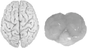
FIgure 19.1
Anatomy of the brain: (a) human and (b) woodpecker.
Search WWH ::

Custom Search