Biomedical Engineering Reference
In-Depth Information
The MR images were segmented using MIMICS (Materialise, Leuven, Belgium) to obtain the
tissue boundaries. For the sake of simplification, the articular cartilages of the bones are fused
with their corresponding bone surfaces in the segmentation process. The boundary surfaces of
the skeletal and skin components were processed using SolidWorks (SolidWorks Corporation,
Massachusetts) to form solid models for each bone and the whole foot surface. The solid model was
then imported and assembled in the FE package, ABAQUS (Simulia, United States).
The FE model of the human foot and ankle consisted of 28 bony segments, including the distal
segments of the tibia and fibula and 26 foot bones, namely, talus, calcaneus, cuboid, navicular,
3 cuneiforms, 5 metatarsals, and 14 components of the phalanges embedded in a volume of encap-
sulated bulk soft tissue (Figure 1.3). The phalangeal bones were connected together and spaced
by 2 mm using solid elastic elements, which represented the thickness of the articulating cartilage
layers and simulated the connection of the cartilage and other connective tissues. The interaction
among the metatarsals, cuneiforms, cuboid, navicular, talus, calcaneus, tibia, and fibula were
defined as contacting elastic bodies to allow the simulation of relative bone movement.
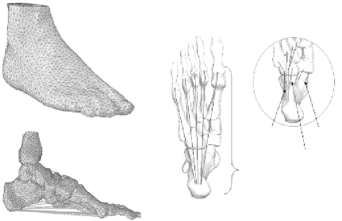
A total of 72 ligaments and the plantar fascia were included and defined by connecting the corre-
sponding attachment points on the bones. Information on the attachment regions of the ligamentous
structures was obtained from the Interactive Foot and Ankle (Primal Picture Ltd., London, 1999).
The attachment points of the major plantar ligamentous structures such as the plantar fascia, long
plantar ligament, short plantar ligament, and spring ligament are depicted in Figure 1.3. The num-
ber of attachment points defined for individual ligaments depended on the width of the ligamentous
structures. For instance, the plantar fascia was divided into five rays of separate sections, linking
the insertions between the calcaneus and the metatarsophalangeal joints (Figure 1.3). The plantar
ligaments and spring ligament were defined by three and two rays of separate sections, while only
a single ray was defined for small ligaments. The attachment points were defined close to the geo-
metrical center of the insertion regions of the ligamentous structures. All the bony and ligamentous
structures were embedded in a volume of soft tissue.
A variety of solids elements in the ABAQUS package can be used to model the foot and
ankle structures. The bony and encapsulated soft tissue structures were meshed into four-noded
Long plantar
ligament
Spring ligament
Short plantar
ligament
Plantar
fascia
FIgure 1.3
The finite element (FE) meshes of the encapsulated soft tissue, bony structures in the lateral
view, and the attachment points of the plantar fascia, spring ligaments, and long and short plantar ligaments
of the FE model.
Search WWH ::

Custom Search