Biomedical Engineering Reference
In-Depth Information
unlikely and suggested that fatigue fracture could be a possible cause of material failure. Another
computational model constructed by Daniel et al. (2006) investigated the effect of necrotic volume
and location. With an increase in necrotic volume and lateralization of the site, the contact pres-
sure within the hip joint increased. These findings suggested a possible source for the subsequent
arthritis.
Grecu et al. (2010) extended the FE model to include the pelvis. With the application of ground
reaction forces and gluteal medius muscle forces, a high strain area was demonstrated on a par-
ticular region of the cartilaginous surface which corresponded to the damaged site of the necrotic
femoral head specimen.
Volokh et al. (2006) simulated seated and walking positions using FE analysis and reported that
buckling was the most important mechanism leading to collapse in ONFH. A parametric analysis
of the cortical thickness and cortical and trabecular elastic modulus was carried out. The buckling
mode was assessed by the critical buckling pressure and predicted pressure. However, Volokh et al.
did not rule out other possible mechanisms, such as fatigue, that could occur concurrently.
10.1.6 i
inVeStiGation
of
tHe
p
atHomecHaniSm
The pathomechanism suggested by various sources in the literature is quite general. Amanatullah,
Strauss, and Di Cesare (2011a) reported that femoral head collapse was basically caused by bone
resorption. After bone resorption, the weakened trabeculae could not accommodate stress concen-
tration and would eventually fracture (Amanatullah, Strauss, and Di Cesare 2011a). In fact, the
bone deterioration in ONFH is quite unique and progresses with different clinical symptoms, and
different patterns of necrosis are speculated to occur in different regions. Figure 10.1 shows some
of the signs of ONFH.
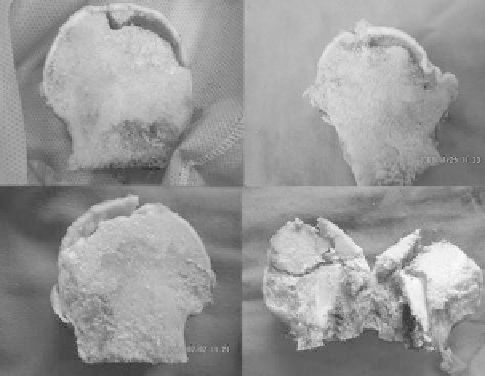
Crescent fractures are the most common findings in ONFH and are illustrated by a crescent shape
on x-ray scans. It is believed that the crescent predisposes the femoral head to collapse, whereby a
portion of the necrotic bone is lost (Figure 10.1b). Fracture of the cortical bone is also seen in some
cases. Although it could result from preceding collapse and unsustainability, a precollapse condition
also reveals the possibility of cortical fracture (Figure 10.1c). A large gray area beneath the necrotic
regions is also seen; this is an area of osteosclerosis. Occasionally, fracture occurs on the boundary
between the necrotic and healthy trabecular bone, as shown in Figure 10.1d.
(a)
(b)
(c)
(d)
FIgure 10.1
(See color insert.)
Typical signs of ONFH: (a) crescent fracture; (b) femoral head collapse;
(c) cortical fracture and osteosclerosis; (d) necrotic boundary fracture.
Search WWH ::

Custom Search