Biomedical Engineering Reference
In-Depth Information
9.4 aPPlICatIonS oF the model
FE analysis of QCT scans is the most advanced method for noninvasive clinical assessment of
femoral strength, and was regarded as biomechanical CT (Keaveny et al., 2010). Identifying the
associations between the subject-specific femoral strength predicted by image-based nonlinear FE
analysis and clinically available information may have the potential for noninvasive clinical assess-
ment of individual hip fracture risk.
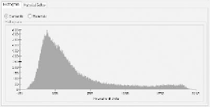
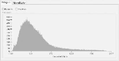
Material distribution can provide more details about the material properties of the proximal
femur than BMD, and it can be easily obtained from QCT data. Figure 9.4 shows the material dis-
tributions of the two models evaluated using MIMICS software, from which it can be clearly seen
that the material distributions of the two models were significantly different, with model A having
more materials with greater elastic moduli.
It is also convenient to obtain the geometrical parameters from QCT data (Lv et al., 2012),
which included neck length (NL), diameter of femoral head (HD), height of femoral head (HH),
offset (OFF), neck shaft angle (NSA), height of the top of the greater trochanter (TRH), thickness
of femur (TOF), and neck diameter (ND). A schematic of the geometric parameters of a proximal
femur (model A) is shown in Figure 9.5. The three-dimensional geometric parameters of the two
models in this chapter are listed in Table 9.2, from which it can be seen that the geometries of the
two models were quite different.
Recently, three independent factors (principal components, or PCs) were extracted from BMD,
material distribution from QCT data, height, weight, and geometric parameters. A high correlation
was found between the three PCs and the strength predicted from FE analysis. Besides BMD, other
parameters, such as material distribution and geometric parameters, also contributed significantly
to femoral strength (Gong et al., 2012).
If developed properly, while taking the most relevant information into consideration, subject-
specific FE modeling has the following advantages: the physical properties of bone measured non-
invasively can derive the nonlinear mechanical properties of the bone; parametric studies can be
performed easily; heterogeneity and anisotropy can be addressed; and the magnitudes and direc-
tions of loading can be varied easily. Accordingly, subject-specific, image-based, nonlinear FE
analysis can quantitatively predict the form and magnitude of loads that may bring about fracture,
as well as the possible location and type of fracture. It offers insight into the fracture mechanism
and may help to predict brittle fracture risk and determine feasible exercises for the elderly to avoid
falls. Subject-specific bone strength and evaluation of the related fracture risk can be obtained non-
invasively based on a patient's clinical information.
(a)
(b)
FIgure 9.4
Material distributions of the two models evaluated using MIMICS software: (a) model A;
(b) model B.
Search WWH ::

Custom Search