Biology Reference
In-Depth Information
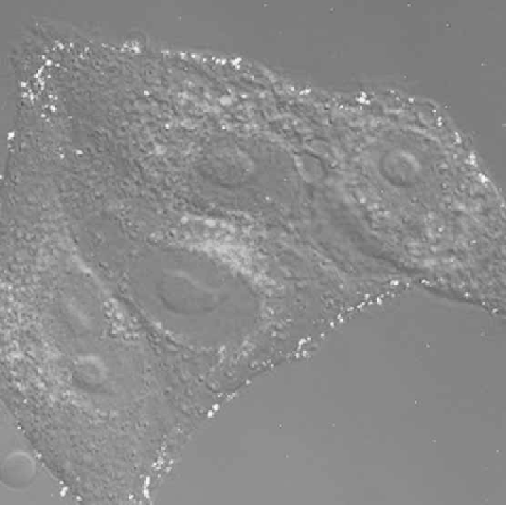
Figure 16.1. Confocal laser scanning microscopy of CD4
HeLa cells incubated with GFP-
tagged HIV-1 virions that are seen as dots in the periphery of the cells. The endosomal compart-
ments are labeled with rhodamine red transferrin and appear mostly in the perinuclear area of the
cells. The cells were ®xed 10 min after application of the virus. The size of the spots corresponding
to the labeled virions does not re¯ect the real size of viral particles. Virions appear much larger
due to the very strong ¯uorescent signal of GFP resulting in scattering of photons in microscope
detector. No colocalization of virions with endosomes could be detected.
revealed signi®cant di¨erences in responses to Tat between di¨erent LTRs. The
authors have found the assay to be accurate and sensitive, but only in a certain
interval of amount of DNA used for transfection. The problem can be alle-
viated and the test can be made easier and more reliable by the generation of
stably transfected cell lines. A stable T-cell line (CEM) expressing a plasmid-
encoding humanized enhanced GFP under the control of HIV-1 LTR was
shown to respond to HIV-1 infection by a 100- to 1000-fold increase in ¯uo-
rescence (Gervaix et al., 1997). The activity of antiretroviral drugs with di¨er-
ent mechanisms of action was evaluated in CEM-GFP cells. All compounds