Biomedical Engineering Reference
In-Depth Information
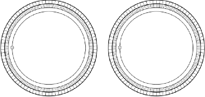
Blank scan
Transmission scan
Patient
Ge-68
rotating rod
source
Figure 2.8: Attenuation correction in PET using a rotating rod source of
68
Ge. Blank and transmission scans are generally acquired before tracer
administration.
scan is acquired to measure the coincidence rate when the patient being im-
aged is in the FOV but has not been given an injection of positron emitter.
Figure 2.8 shows a schematic for measured attenuation correction using a ro-
tating rod source of positron emitter
68
Ge. Attenuation correction factors are
then determined by calculating the pixelwise ratio of the measured projection
data obtained from the blank scan and the transmission scan. The major draw-
back of this approach is that statistical noise in the transmission data would
propagate into the emission images [46, 47]. Therefore, transmission scans of
sufficiently long duration have to be acquired to limit the effect of noise propa-
gation. Depending on the radioactivity present in the external radiation source
and on the dimension and composition of the body, transmission scans of 15-30
min are performed to minimize the propagation of noise into the emission data
through attenuation correction, at the expense of patient throughput. Further,
lengthened scan duration increases the likelihood of patient movement, which
can cause significant artifacts in the attenuation factors for particular LoRs.
Application of analytical, so-called calculated attenuation correction elim-
inates the need for a transmission scan, thus making this method attractive
in many clinical PET centers. This method assumes uniform skull thickness
and constant attenuation in the brain and skull. However, such assumptions