Biomedical Engineering Reference
In-Depth Information
2.7 Detectors
To image the distribution of positron-emitting isotope in the body, both of the 511
keV photons emitted from positron annihilation must be detected in coincidence.
Unlike other instruments used in nuclear medicine, PET uses electronic rather
than lead collimators to detect signal (event) results from annihilation of the
positron and an electron. The probability of detecting both photons depends
on the detector efficiency, which is strongly related to the stopping power of
the scintillator and the thickness of the scintillator used in the detector. Early
generation of PET scanners used NaI(Tl) crystals, the same material used in
gamma camera. Modern PET scanners use much denser scintillators, such as
bismuth germanate oxide (BGO) [27], which has been the scintillator of choice
for more than two decades due to its very high density and stopping power for
the 511 keV gamma rays. In order to provide higher detection efficiency and
spatial resolution with lower production cost, a number of detector designs
were proposed in the 1980s and the most successful one was the
block detector
technique proposed by Casey and Nutt, using BGO crystal [28]. A typical BGO
block detector comprises a rectangular block consisting of between 6
×
8 and
8
×
8 individual scintillation crystals, attached to an array (usually 2
×
2) of
photomultiplier tubes (PMTs) at which the scintillation light is amplified and
converted into electrical signal for the coincidence detection circuit to register.
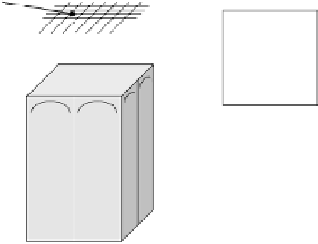
A schematic outline of such a block detector is shown in Fig. 2.3. The BGO
element in which a gamma ray interacts is determined by the relative light output
Scintillator
array
P
1
+P
2
-P
3
-P
4
P
1
+P
2
+P
3
+P
4
X=
PMTs
P
1
-P
2
+P
3
-P
4
P
1
+P
2
+P
3
+P
4
Y=
Figure 2.3: Schematic diagram of a BGO block detector commonly used in
commercial PET systems.