Biomedical Engineering Reference
In-Depth Information
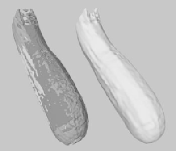
Figure 8.10: Three low-resolution MR scans of a zucchini that have been in-
dividually colored and overlaid to demonstrate their imperfect alignment. The
level set model on the right is derived from the three low-resolution scans.
8.5 Segmentation of DT-MRI Brain Data
Diffusion tensor magnetic resonance imaging [55, 56] (DT-MRI) is a tech-
nique used to measure the diffusion properties of water molecules in tissues.
Anisotropic diffusion can be described by the equation
∂
C
∂
t
=∇·
(
d
∇
C
)
,
(8.25)
where
C
is the concentration of water molecules and
d
is a diffusion coefficient,
which is a symmetric second-order tensor
⎛
⎝
⎞
⎠
.
D
xx
D
xy
D
xz
D
yx
D
yy
D
yz
D
zx
D
zy
D
zz
d
=
(8.26)
Figure 8.11 presents a “slice” of the diffusion tensor volume data of human brain
used in our study. Each subimage presents the scalar values of the associated
diffusion tensor component for one slice of the dataset.
Tissue segmentation and classification based on DT-MRI offers several ad-
vantages over conventional MRI, since diffusion data contains additional phys-
ical information about the internal structure of the tissue being scanned. How-
ever, segmentation and visualization using diffusion data is not entirely straight-
forward. First of all, the diffusion matrix itself is not invariant with respect to
rotations, and the elements that form the matrix will be different for different