Biomedical Engineering Reference
In-Depth Information
the “spines” that cover this dendrite. These types of “judgments” that humans
make when they perform such tasks by hand are a mixed blessing. Humans can
use high-level knowledge about the problem to fill in where the data is weak,
but the expectations of a trained operator can interfere with seeing unexpected
or unusual features in the data.
Figure 8.8(c) presents models from four samples of an MR series of a devel-
oping frog embryo. The top left image (hour 9) shows the first evident structure,
the blastocoel, in blue, surrounded by the outside casing of the embryo in gray.
(b)
(a)
(c)
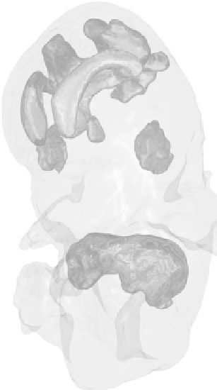
Figure 8.8:
(a) Final mouse embryo model with skin (gray), liver (blue), brain
ventricles (red), and eyes (green). (b) Hour 16 dataset. (c) Geometric structures
extracted from MRI scans of a developing frog embryo, with blastocoel (blue),
blastoporal lip (red), and archenteron (green). Hour 9 (top left), hour 16 (top
right), hour 20 (bottom left), and hour 30 (bottom right).