Biomedical Engineering Reference
In-Depth Information
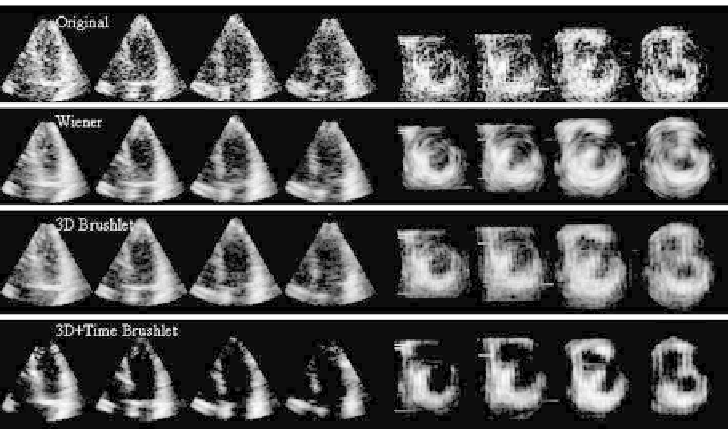
Figure 6.18: Spatio-temporal denoising with brushlet expansion on RT3D ul-
trasound data illustrated on four long-axis and four short-axis slices.
denoising and Wiener filtering on RT3D ultrasound data. Adding the time dimen-
sion leads to images with better contrast and sharper contours while preserving
the original textural aspect of the ultrasound data. Wiener filtering provided
good results but introduced blurring artifacts that severely altered the quality
of the short-axis denoised images. This type of artifact is unacceptable in medi-
cal applications where anatomical structure detail needs to be preserved. It was
also observed that the epicardium borders were enhanced with sharper contrast
when combining brushlet spatial and temporal denoising. Such enhancement is
very desirable for quantification of LV mass and wall thickness analysis that re-
quires segmentation of both the myocardial endocardial and epicardial borders.
6.3.5.2 Cross-Scale Regularization for Tomographic Images [60]
Tomographic image modalities such as PET and SPECT rely on an instable in-
verse problem of spatial signal reconstruction from sampled line projections.
Tomographic reconstruction includes backprojection of the sinogram signal via
Radon transform and regularization for removal of noisy artifacts. Because the
Radon transform is a smoothing process, backprojection in the presence of
additive noise is an ill-posed inverse problem that requires a regularization of
the reconstructed noise component, which can become very large. Standard