Biomedical Engineering Reference
In-Depth Information
Original
2D Denoising
3D Denoising
(a)
Original
2D Denoising
3D Denoising
(b)
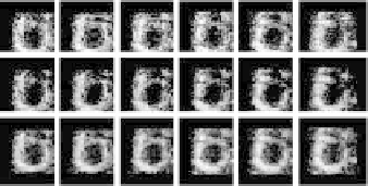
Figure 6.13: 2D versus 3D spatial denoising on RT3D ultrasound data. (a) Series
of six consecutive short-axis slices extracted from a clinical data set. (b) Series
of six consecutive long-axis slices extracted from the same clinical data set.
wall on a black background. The size of a single volume was 64
×
64
×
64 and
there were 16 frames growing in time. The volume increased by 70% over 16
time frames, similar to the average ejection fraction in normal patients.
The phantom was corrupted with two types of noise: (1) multiplicative
speckle noise with uniform distribution and (2) multiplicative speckle noise
with Rayleigh distribution.
The level of speckle noise was set so that the signal-to-noise ratio (SNR)
of the noisy data was equal to
−
15 dB. Cross-sectional slices through a single
volume of the noisy phantoms are displayed in Fig. 6.15.
Time
1
8
16
Figure 6.14:
Mathematical phantom. Ovoid volume with 16 frames growing in
time.