Biomedical Engineering Reference
In-Depth Information
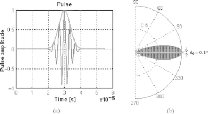
Figure 1.9: Typical ultrasound pulse and its Gaussian envelope (a). The trans-
ducer pattern radiation (b).
Eq. (1.2) in the transducer coordinate system is based on a discrete representa-
tion of the tissue of individual scatterer elements with given position and DBC
with respect to the transducer coordinates given by:
N
σ
i
(
R
,,
Z
)
|
R
i
|
S
(
R
,,
Z
,
t
,δ
)
=
C
o
ζ
(
t
,δ
)
(1.5)
i
=
1
where
δ
is given by Eq. (1.1), and
ζ
(
t
,δ
) is the impulse function given by
Eq. (1.4). If we consider only the axial intensity contributions,
C
o
can be written
as [14]:
C
o
=
ρ
o
ck
2
2
o
A
v
(1.6)
8
π
where
A
is the transducer area.
1.4 Principal Features of IVUS Data
1.4.1 Image Resolution
Resolution is the capacity of a technique or an instrument to separate two events
or objects in time and/or space [14]. At the moment, much of the effort in the
design of new transducers is centered in improving the spatial and the tempo-
ral resolution. Unfortunately, most of the medical applications demand that the