Biomedical Engineering Reference
In-Depth Information
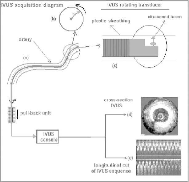
Figure 1.3: The IVUS catheter is manually positioned within the artery (a)
and extracted by a pull-back unit at a constant linear velocity and rotated at
a constant angular velocity. The information is transformed by the IVUS console
as a unique cross-section artery gray-levels image (d) or a longitudinal image
sequence (e).
and constant angular velocity of 1800 rev/min. The pivoting transducer sends a
radially focused beam of ultrasound and receives its corresponding echoes. The
radial lines obtained for different transducer angular positions are adequately
processed, giving, as a result, a 2D cross-section artery image (Fig. 1.3(d)). The
sequence can be shown as a longitudinal sequence, which gives a longitudinal
artery cut (Fig. 1.3(e)). The resolution of an ultrasound image is directly related
to the ultrasound signal frequency: high frequencies allow one to obtain better
resolution. Nevertheless, when the frequency is increased, the attenuation of
the waves of ultrasound increases while penetrating the biological tissue. The
typical frequencies in the IVUS technique are in the rank of 20-50 MHz, with
inferior resolutions of 50
µ
m.