Biomedical Engineering Reference
In-Depth Information
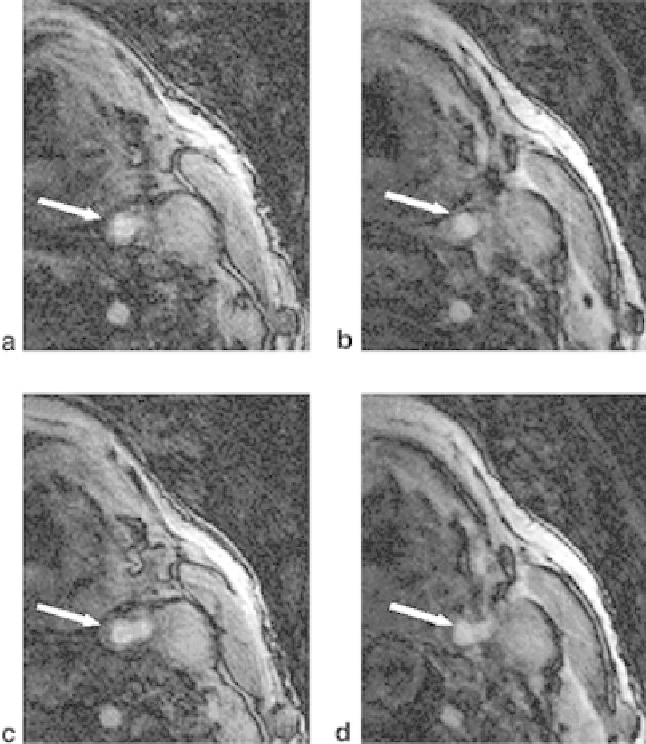
Figure 3.31:
Source images of the 3D TOF MRA of the left carotid artery of a
volunteer: (a and b) inferior and (c and d) at the carotid bifurcation. Images were
acquired (a and c) without VTE and (b and d) with VTE (16 TE segments). The
imaging parameters were as follows: matrix
=
256 A
∼
256 A
∼
32, slice thickness
=
1 mm, TR
=
24 ms, FOV
=
14 cm, and TE
=
1.8/2.9 ms for VTE on/off. MT was
not applied. The reduced signal indicated by arrows in a and c was much more
uniform in images b and d with VTE.
bolus of Gadoteridol, and a 3D pulse sequence with a 66% sampling efficiency.
This spatial resolution allowed visualization of intracranial aneurysms, carotid
dissections, and atherosclerotic disease including ulcerations. Potential draw-
backs of 3.0 T MRA are increased SAR and T(*)2 dephasing compared to 1.5 T.