Biomedical Engineering Reference
In-Depth Information
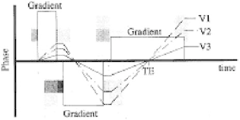
Figure 3.26:
V1, V2, and V3 effect.
3.2.11 Black Blood MRA
Black blood MRA is another technique for MRA in which flowing blood appears
dark rather than bright. It appears as negative of bright blood MRA. Rapidly
flowing blood in arteries exhibits the TOF MRA signal losses. Slow flowing
blood in veins appears as higher signal intensity. Various flow presaturation
pulses and dephasing methods via gradients are employed in this technique to
render flowing blood as black. This technique uses the MIP algorithm. Black
blood MRA has several advantages. They offer no overestimation for the degree
of stenosis and no dephasing in vessel turns that mimic stenosis. On the other
hand, the technique has disadvantages that calcified plaque appears dark. Thus,
this technique may underestimate the degree of stenosis or invisible plaques.
Other black materials such as air or bone may mimic the blood flow.
Black blood MR angiograms make use of another time-of-flight
phenomenon—the signal void observed for flowing blood in spin echo images.
Unlike white blood, or INFLOW angiograms, which use a gradient echo sequence
to enhance flowing blood and saturate static tissue, black blood angiography
uses a spin echo sequence with presaturation to increase the signal of the tissue
and to create a signal void (i.e. no MR signal) for flowing blood. The data is then
processed using an MIP algorithm to yield the final MR angiogram. Black blood
magnetic resonance angiography offers the advantage that signal voids due to
turbulent flow are avoided. However, the contrast between vessel and static
tissue may be lower, arterial and venous flow cannot be easily distinguished,
and several regions of signal void such as nasal sinuses exist on images. Despite
these disadvantages, black blood MRA may prove useful in the determination
of some pathologies, such as severe stenotic lesions.