Biomedical Engineering Reference
In-Depth Information
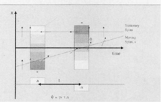
Figure 3.15: Flow is encoded in one direction using bipolar gradients. Through
encoding, stationary tissue receives zero phase shift,
φ
, while moving spins
receive a phase shift proportional to their velocity,
v
.
second gradient in the opposite direction. Flowing spins change the position
during application of the dephasing and rephasing gradients (see Fig. 3.16). As
in PC MRA, both magnitude and phase images can be obtained with information
on direction of flow in the vessel (see Fig. 3.17).
The effect of velocity-dephasing on phase information indicates the flow di-
rection in right-left (R/L), superior-inferior (S/I), or anterior-posterior (A/P).
This effect may be described as follows: flow-induced different phase shifts are
generated due to spins moving in-plane along frequency gradient in different
directions (see Fig. 3.18 shown by zig-zag arrows). Conventionally, spin flow is
higher at the center than near the wall due to laminar flow. Due to this differ-
ence, spins at the center cause larger phase shift than the phase shift by slower
peripheral coherence. This results in velocity dephasing and total signal loss
called “flow void.” In this way, phase information is transferred to a magnitude
contrast.
3.2.3.1 2D Phase Contrast Angiography
The primary advantage is that a variety of velocity encoding may be opted in a
short period of time (within a few minutes). If limited angiographic information