Biomedical Engineering Reference
In-Depth Information
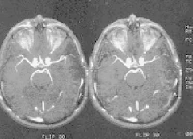
Figure 3.13: Effect of flip angle in 3D TOF angiogram images shows at different
flip angles 20
◦
(left) and 30
◦
(right).
therefore maximizes intravascular signal. Intraluminal signal loss may still occur
in spite of the use of first-order motion compensation. High order motions such
as jerks and acceleration may still produce regions of signal loss due to the phase
dispersion. For instance, blood flow in carotid siphon experiences centripetal
acceleration along the carotid vessel's outer wall. In 3D TOF images, the effects
of acceleration are not compensated and result in linear regions of signal loss
at curves in the carotid artery and proximal middle cerebral artery. However,
magnetic susceptibility effects from the adjacent paranasal sinuses play a minor
role in the loss of signal intensity in the juxtasellar carotid artery and proximal
middle carotid artery at short TE. Mostly, signal intensity losses are observed at
the bends of these arteries during diastole. Higher order motion compensation
gradients extend TE. At extended TE, susceptibility effects are significant and
the signal loss is more apparent.
3.2.2.1.6 Slice Thickness.
Slice thickness also contributes to signal loss.
Thicker slices show significant signal loss. Thin slices exhibit phase dispersion
within the voxel which minimizes signal loss and effects of intravoxel dephasing.
However, thin slices reduce signal-to-noise ratio and the volume of interest.
Other important refinements in this technique are described in Section 3.4.
3.2.3 Phase Contrast MRA
Phase contrast (PC) MRA is based on the fact that the phase gain of flowing blood
through a gradient is proportional to its velocity (assuming constant velocity).