Biomedical Engineering Reference
In-Depth Information
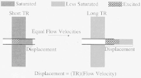
Figure 3.7: Effect of TR is shown. At short TR, stationary tissue is partially
saturated causing saturated blood flowing out of slice and replaced with unsat-
urated blood appearing as high signal in the blood relative to surrounding tissue
(on left). At long TR, stationary tissue may recover between excitations and
excited blood flows out of the slice before it is refocused to form echo or high
signal of stationary tissue with no signal from flow.
image contrast in 2D TOF images include flow velocity and direction, vessel
geometry, T1 of blood and stationary tissues, flip angle, TE, and slice thickness.
3.2.1.1.2 Flow Velocity.
Initially, flow-related enhancement increases with
the flow velocity. At moderate flow velocity rates, there is flow-related enhance-
ment for a complete new set of the spins. Later, no further increase is possible
in image contrast or signal intensity.
3.2.1.1.3 Vessel Geometry.
The orientation of the blood vessel to the slice
plane also affects vascular signal intensity. Maximum inflow enhancement oc-
curs when blood flow is perpendicular to the imaging plane. When a vessel
travels obliquely through the slice or the vessel lies within the slice plane, the
flowing spins are subjected to multiple RF pulses. As a result, spins begin to
become saturated. As a result, intravascular signal intensity decreases and the
vessel may be incompletely visualized.
3.2.1.1.4 Slice Thickness.
The thinnest slices maximize inflow enhance-
ment. These thin slices reduce the effects of in-plane flow. Typically, for our
carotid bifurcation imaging, a nominal 1.5 mm slice is obtained using a narrow
bandwidth RF pulse at 625 Hz and gradient amplitudes of 1 G/cm or 10 mT/m.