Biomedical Engineering Reference
In-Depth Information
from the uncompensated sequence or by subtracting a fully presaturated image
from a unilaterally presaturated image. The image acquisition in the interleaved
fashion will further minimize the motion artifacts.
3.1.2.3 Spin Phase Phenomenon
This effect is based on the motion in a vessel in the direction of magnetic field
gradients. It leads to the precession phases different from zero in bulk motion,
while the magnitude of the magnetization vector remains unaffected. All of the
moving spin isochromats within the voxel experience the same phase change.
Interestingly, the moving fluid will have a different phase. Flowing blood gives
rise to a velocity profile in a vessel, divided into different voxels. Due to phase
change along the vessel wall and surrounding regions, velocity variation is ob-
served due to phase changes either 90
◦
or 180
◦
. It causes considerable signal
loss in the voxel at the location of fat tissue.
Suppose a velocity difference of 1 cm/sec within a voxel produces preces-
sional phase changes of approximately 360
◦
, it will lead to complete signal loss
by use of SE sequence with typical gradient values. For slower blood flow, in-
travascular signal is seen less dephased and is more prominent at the center of
the vessel such as accelerated blood flow. With acceleration, the signal loss that
results from the dephasing of spin isochromats increases in proportion to the
echo number (see Fig. 3.4). For constant velocity motion, this method may be
known as even-echo rephrasing or even-echo refocusing for the flow along the
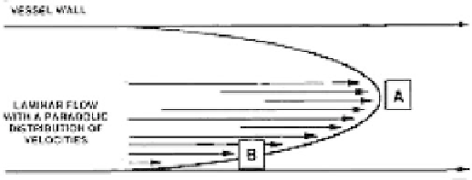
Figure 3.4: Intravoxel spin-phase dispersion due to incoherence is shown near
the center of the vessel (point A) for minimal phase dispersion. Point B near
the vessel wall encompasses a large range of velocities resulting with intravoxel
dephasing and signal loss.