Biology Reference
In-Depth Information
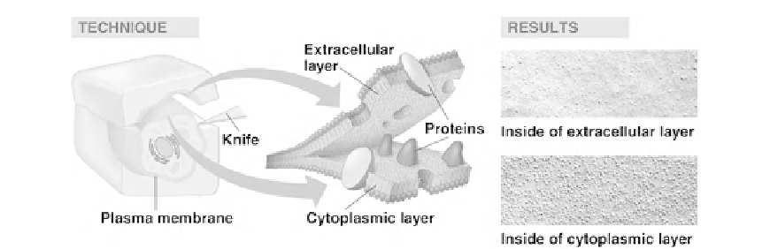
FIGURE 11.5
The technique of freeze fracture electron microscopy. The process is briefly discussed in the text
and is discussed in much more detail in a 2007 review paper by Severs
[20]
. Note there are many more proteins
projecting from the cytoplasmic leaflet than from the extracellular leaflet
[21]
.
of the membrane (
Figure 11.5
)
[21]
. The integral membrane proteins appear as particles of
about 80 to 100
˚
in diameter. Usually they are randomly dispersed, but may be arranged
in groups. Some particles are preferentially associated with one membrane leaflet face or
the other (as demonstrated in
Figure 11.5
). Freeze fracture EM supplies direct evidence that
proteins reside inside membranes and are not just stuck on the surface. The particle distribu-
tion also indicates that there is no long-range order, instead observed order is only a few tenths
of a micron and the membrane bilayer sea is indeed very crowded
[22]
.
Examples of long-range order due to cytoskeletal attachments have been observed. In an
early report from 1976, Yu and Branton
[23]
showed that egg lecithin liposomes have very
smooth membrane leaflet surfaces that were devoid of protein particles. When the erythro-
cyte protein band 3 was reconstituted into the liposome membranes, dispersed particles
appeared in freeze fracture that closely resembled what was imaged for natural erythrocyte
membranes. When the pH was dropped to 5.5, the particles observed in the erythrocyte
clumped together due to action of the cytoskeleton. Interestingly, when spectrin and actin,
components of the cytoskeleton, were added to the reconstituted band 3-egg lecithin lipo-
somes and the pH was decreased to 5.5, particle clumping was also observed.
Unfortunately, freeze fracture EM can be fraught with potential artifacts. Just the process
of rapid freezing in liquid nitrogen produces destructive ice crystals. Therefore pretreatment
with a cryoprotectant (glycerol) and glutereraldehyde is often employed. The biological
membrane itself is not directly imaged. Instead a replica is made by shadowing the sample
with platinum at a 45
angle to highlight the topography. The replica is further strengthened
with a thin layer of carbon. The replica is then carefully washed off the sample and imaged by
EM. Every additional step in a complex process is a potential source of artifacts.
C. IMAGING OF MEMBRANE DOMAINS
1. Macrodomains
The term 'domain' has come to be used extensively in the life sciences literature. Unfortu-
nately, 'domain' means quite different things to different investigators. Funk and Wagnalls'