Biology Reference
In-Depth Information
molecular locations and interrelationships would be altered. Therefore membranes must
have both static and dynamic components. While static describes what is there, dynamics
describes how the components interact to generate biological function.
Every cell in the human body is a tightly packed package of countless membranes. The
human body is composed of ~63 trillion cells (6.3
10
13
cells), each of which is very small.
For example, a typical liver cell would have to be 5X larger to be seen as a speck by someone
with excellent vision (it is microscopic). Each liver cell has countless numbers of internal
membranes. If you could somehow open one single liver cell and remove all of the internal
membranes and sew them together into a quilt, the quilt would cover ~840 acres, the size of
New York's Central Park! And that is from one single cell. Therefore, there are enough
membranes in a human body (6.3
10
13
cells) to cover the earth millions of times over!
All life on Earth is far more similar than it is different. Living organisms share a number of
essential biochemical properties, collectively termed the 'thread of life'. Included in these
essential properties is ownership of a surrounding plasma membrane that separates the cell's
interior from its external environment. It is likely that all living things inhabiting planet Earth
today arose from a single common ancestor more than 3.5 billion years ago. The first cell
probably contained minimally a primitive catalyst (a pre-protein), a primitive information
storage system (a pre-nucleic acid), a source of carbon (perhaps a primitive carbohydrate)
and this mixture had to be surrounded by a primitive plasma membrane that was likely
made of polar lipids. Membranes were therefore an essential component of every cell that
is alive today or has ever been alive.
With 3.5 billion years of biological evolution, the complexity of membranes in cells has
greatly expanded from that of a simple surrounding plasma membrane to where they
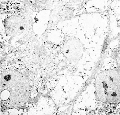
now occupy a large portion of a eukaryote's interior space. An electron microscopic
picture of a 'typical' eukaryotic (liver) cell is shown in
Figure 1.1 [1]
.Itisevidentfrom
the complexity of this micrograph that identifying, isolating, and studying membranes
will be a difficult task.
FIGURE 1.1
Transmission electron micrograph of a liver cell, a 'typical' cell
[1]
.