Biology Reference
In-Depth Information
FIGURE 9.6
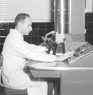
Humberto Fernandez-Moran (1924
e
1999)
[56]
.
for example, cannot be obtained by X-ray diffraction. Note that the addition of cholesterol
(dotted line) increases the distance between the electron dense peaks demonstrating that
cholesterol increases membrane bilayer thickness. Therefore, X-ray diffraction does not yield
molecular detail of membrane structure, but does support the concept of a lipid bilayer.
C. MEMBRANE ASYMMETRY
Even before membrane asymmetry had been experimentally proven, it was universally
agreed that proteins and probably carbohydrates should be 100% asymmetrically distributed
across the membrane. The situation for membrane lipids, however, was not as certain.
Protein Asymmetry
It seems logical that all functional membrane proteins should be oriented in only a single
trans-membrane direction to prevent futile cycles. For example, if the function of a protein
was to pump a solute into a cell, it would not make any sense if some of the same solute trans-
port proteins were oriented in the opposite direction, thus pumping the same solute back out
of the cell. But logic is not proof. A classic 1964 electron microscopy (EM) paper by Humberto
Fernandez-Moran
(Figure 9.6)
is often cited as the first definitive evidence that membrane
proteins are 100% asymmetrically distributed across membranes. Fernandez-Moran was
a pioneer in electron microscopy and has been credited for developing the diamond knife
and ultra-microtome for EM use. Most amazing is the fact that he was able to obtain his
Ph.D. from the University of Munich in 1944! In 1964 he published a paper with David Green
who later proposed the Lipoprotein Subunit model for membranes (Chapter 8). In this paper
Fernandez-Moran used negative staining with phosphotungstate to obtain EM images of what
was then known as a mitochondrial elementary particle (EP)
[5]
. We now know EP is actually