Chemistry Reference
In-Depth Information
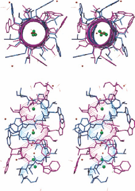
Figure 1.13 Stereo relaxed eye view of the crystal structures of one channel of gramicidin A
dimer (top, viewed through the channel; bottom, side view; PDB ID 1AV2). Cs
þ
ions located
in the channel are shown in green spheres, water molecules are shown in small red spheres.
this obstacle, microorganisms excrete Fe
3þ
-specific siderophores which bind Fe
3þ
with
extraordinarily high affinity constants in the range of
10
30
-10
52
M
1
and transport Fe
3þ
into cells via specific receptors [156,171]. There are three families of siderophores, differ-
ing from each other by their iron-binding sites: hydroximate- (such as ferrichrome from
Penicillium
and the edible
Ustilago
), catechol-containing (e.g., enterobactin from
E. coli
),
carboxylate-containing (like the simple citrate), and their combinations (such as aerobac-
tin) [172]. Upon Fe
3þ
binding, the ferrichromes fold to afford an octahedral metal coordi-
nation sphere with a more compact conformation than their metal-free apo-forms
(Figure 1.14, top). The complexes are recognized and transported into the cells by spe-
cies-specific receptors. For example, although ferrichrome A serves as an iron carrier for
fungi, it does not in bacteria [173]. The folding is also stereo-specific. While the iron
complexes of ferrioxamines B [174], D
1
[175], and E [176] and desferrioxamine E [177]
fold into a mixture of L- and D-
cis
isomers, ferrichrome complexes [178] are exclusively
L-
cis
isomers. A few structures of the transporter protein FhuA (ferric hydroxamate
Search WWH ::

Custom Search