Chemistry Reference
In-Depth Information
found to be around 30% after 24 h. Furthermore, helicates with Sm
III
,Tb
III
and Yb
III
behave in the same way as [Eu
2
(L7)
3
]. Since the luminescence lifetimes are different,
some time-based discrimination is feasible, in addition to wavelength discrimination [77].
The helicate stains have to be excited at around 320 nm, which is a wavelength at which
cell damage may be induced. Additionally, confocal microscopes usually have excitation
wavelengths
405 nm. For this reason, H
2
L11 (Scheme 6.2) was developed, with the
hope of shifting the excitation wavelength more towards the visible range. Indeed, with
respect to (L7)
2
, the absorption maximum of (L11)
2
is red-shifted by 28 nm, from 322
to 350 nm; and another, slightly weaker absorption band occurs at 370 nm (Figure 6.8,
left). This results in brighter confocal luminescence microscopy images, as shown on the
right side of Figure 6.8, while maintaining the same biochemical behaviour (cytotoxicity,
internalization mechanism, localization, egress) as the helicate with (L7)
2
.
Another and potentially better way of shifting the excitation wavelength is to excite the
lanthanoid bioprobes via two- or three-photon processes. The excitation wavelength can
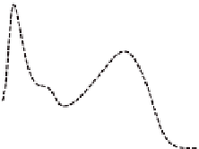
(a)
180
[Eu
2
(L11)
3
]
160
140
120
100
[Eu
2
(L7)
3
]
2
(
3
]
[
80
60
40
20
0
200
250
300
350
400
λ
/nm
(b)
100
[Eu
2
(L11)
3
[Eu
2
(L7)
3
]
90
80
70
60
50
40
0
100
200
300
400
500
c /
μ
M
Figure 6.8 (a) Absorption spectra of [Eu
2
(L
i
)
3
](
i
¼7, 11) in aqueous solution at pH 7.4 (Tris-
HCl 0.1M). (b) Integrated luminescence intensity from confocal microscopy images of HeLa
cells incubated at 37
C with various concentrations of [Eu
2
(L7)
3
] and [Eu
2
(L11)
3
].
Reprinted
with permission from [50]. Copyright 2009 WILEY-VCH Verlag GmbH & Co. KGaA,
Weinheim.


Search WWH ::

Custom Search