Biomedical Engineering Reference
In-Depth Information
studies Ca-aluminate material was compared to the PMMA-material CMW 1, and in the
sheep study Ca-aluminate material was compared to the PMMA-material Vertebroplastic,
and to the Bis-GMA material Cortoss
TM
. The results are summarized in Table 9.
Implantation studies
Species
Reference material
Result
6-week femur
Rabbit
CMW-1
Minimal inflammation,
very few inflammatory
cells were present in bone,
bone marrow and adipose
tissues.
6 (12) -month femur
Rabbit
CMW-1
No Al- accumulation
No inflammation,
no Al-accumulation
Table 9. Implantation studies in femur rabbit, and in vertebrae sheep, details in
(Hermansson et al 2008)
12-week vertebrae
Sheep
Vertebroplastic
Cortoss
The 6 months femur study in rabbits included a 12 months subgroup. The amount of
aluminium in blood and selected organs was analysed. The main target organs of the
animals (kidney, lung, liver) were histopathologically investigated. Granulomatous
inflammation in the cavity, pigmented macrophages and new bone formation were the
treatment-related observations at 6- and12-months examination. No difference between Ca-
aluminate material and PMMA was detected. There were no signs of aluminium
accumulation in the analysed tissues.
In the 12-week study, the histopathology of vertebrae obtained one week after surgery
showed the most severe inflammatory reaction to the surgery in the
sham
operated
vertebrae. The bone marrow in the vertebrae filled with Ca-aluminate was not reported to
be infiltrated by any inflammatory cells. In vertebrae obtained 12 weeks after surgery no
inflammatory reactions were reported, and no obvious differences were observed in the
pathological reactions to the surgery (sham) or the filler materials. Overview of the
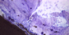
histological contact zone to the Ca-aluminate based material is shown in Figure 4.
The analysis of serum samples showed low concentrations of aluminum in comparison to
what is normal in humans. Since the concentration of aluminum did not increase after
surgery and in some instances was lower after surgery than in the 0-samples, one may
regard these concentrations as within the normal variation.
Fig. 4. Histology image of an experimental Ca-aluminate material (black) in close contact
with sheep vertebral bone.


