Biomedical Engineering Reference
In-Depth Information
The patient was stable from clinical and infective point of view, confirmed by hematologic
exams; so that we decided to underwent the patient to biomaterial infiltration with
Polyacrylamide. The sites of treatment were the areas where the atrophy and the
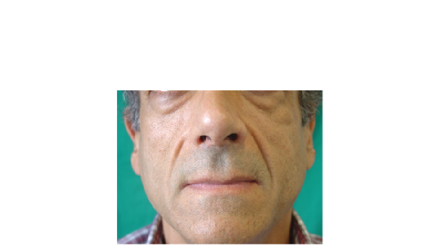
lypodistrophy are happened. Clinical-aesthetic and infective results in six years of follow-up
were good. (FIGURE 4)
Fig. 4. Particular of the the lypodistrophy area after treatment
Polyalkylimide - Clinical case 4:
D.L.B, a male of 30 years old, showed an atrophy of the
middle and inferior third of the face. The patient eight years before, referred a not treated
facial trauma. The objective exam showed the presence of a moderate atrophy of the rigth
hemiface, associated to aesthetic and functional alterations. In particular, a combination of
the right orbital-malar asymmetry complex, and a rigth orbital oenophtalmo was noticed as
well. 7. The patient referred about the appearance of dyplopia. It has not been well
clarifyied. The patient underwent before further clinical and radiological checks exams to
study the soft tissues, and to evaluate the ocular motility, so as a Perry Romberg Syndrome
was suggested for diagnosis. A surgical treatment was planned to correct the oenophtalmo
and to restore the ocular motility correcting the dyplopia disfunction. On the other hand,
this surgery has been performed with the goal of resolving the atrophy and the face's
deformity through the use of porous polyetilene. Afterwards, a treatment of polyackylimide
infiltrations has been planned. The patient, after 1 week from the treatment with
biomaterial, presented a good tolerance and a total restoration of the facial eurytmia. An
ultrasonographic evaluation was performed to value the integration of the biomaterial after
6,12 and 24 months; this exam showed a compartment of the biomaterial implanted,
associated to fibrotyc branches compatible with the basal pathology.
Porous polyethylene - Clinical Case 5:
S.B, a female of 40 years old, referred facial
asymmetry. The objective exam showed a skletal deformity of mandibular and maxillary
component, associated to mycrogenia
.
(FIGURE 5)
The patient presented a congenital nose deformity too. The surgical treatment was planned
with mandibular promotion by means graft of porous polietylene. Then in the same surgical
step a graft “on lay” of septal cartilage was positionated on the nasal dorsum.
Ultrasonographic checks after 1 month, 23 months and 6 months were optimal; radiologic
exams were performed to check the planted biomaterial. These investigations verified a
good tolerance and a well fixed of the biomaterial
.
(FIGURE 6)