Biomedical Engineering Reference
In-Depth Information
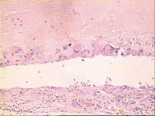
Fig. 7. Inflammatory cell infiltrate is visible 6 weeks after operation. Histocytes gradually
grow deep into the scaffold. No signs of tissue necrosis are found. (×200)
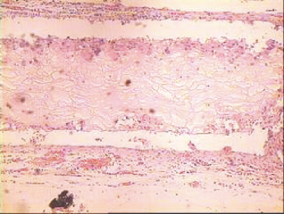
Fig. 8. There are a few parts with eosinophilic staining inside the material disappears 6
weeks postoperatively, which shows disaggregation phenomenon, while it's general
structure remains. (×200)