Biomedical Engineering Reference
In-Depth Information
The UV-photopolymerization via the acrylate end-groups on the sP(EO-stat-PO) arms does
not only allow topographic patterning of the hydrogel's surface, but also enables tuning of the
crosslinking density, hence stiffness. Thus, varying the amount of added photoinitiator (PI)
and crosslinker (CL) represents a practical approach to controlling the mechanical properties
of PEG-based hydrogels (
Figure 3
). This is of high relevance for biomedical applications, as it
is well known that cells feel and respond to the stiffness of the underlying substrate (Discher et
al., 2005; Engler et al., 2006; Yeung et al., 2005). PEG-based hydrogels with distinctly different
mechanical properties were fabricated; the resulting hydrogels from 3 different formulations
are denoted as soft, intermediate and stiff (Schulte et al., 2010; Diez et al., 2011).
The stiffness in the swollen state, which is obviously the most relevant for cell culture, was
shown to be approximately half of that measured in the dry state, ranging from ~100 kPa for
the softest to 1 MPa for the stiffest, thus covering one order of magnitude in elastic modulus
(Figure 3).
2.2 Cytotoxicity assessment of PEG-based substrates
As the material has not been used in this exact composition before, cytotoxicity tests were
conducted to prove that possible traces of unreacted acrylate end-groups, photoinitiator or
crosslinker did not interfere with cell viability. The impact of the material on the viability of
L929 fibroblasts was tested with an indirect cell test. Cell membrane integrity as an
important indicator for cell viability was tested with a commercially available enzymatic
assay. Values shown in
Figure 4
were derived by comparison with a control sample of
mortalized cells (incubation with DMSO), which was set to 100 % cytotoxicity. It should be
noted that no direct test was possible as PEG is known to be anti-adhesive and the majority
of the seeded cells would not stay attached to the PEG-substrate and could therefore not be
used for quantitative studies.
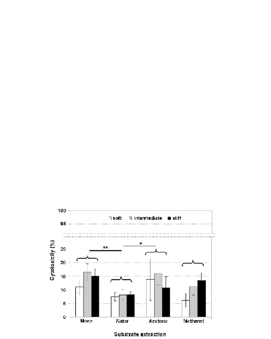
Fig. 4. Cytotoxicity of bulk PEG samples; shown are the results of an indirect cytotoxicity
test with L929 cultured for 24 h in PEG conditioned (72 h) medium. The PEG samples had
before been extracted in water, acetone or methanol for 24 h. The test was performed in
triplicate with substrates of three different crosslinking densities. Statistical significance
indicated by **: p<0.01; *: p<0.05.