Biomedical Engineering Reference
In-Depth Information
3. Results and discussion
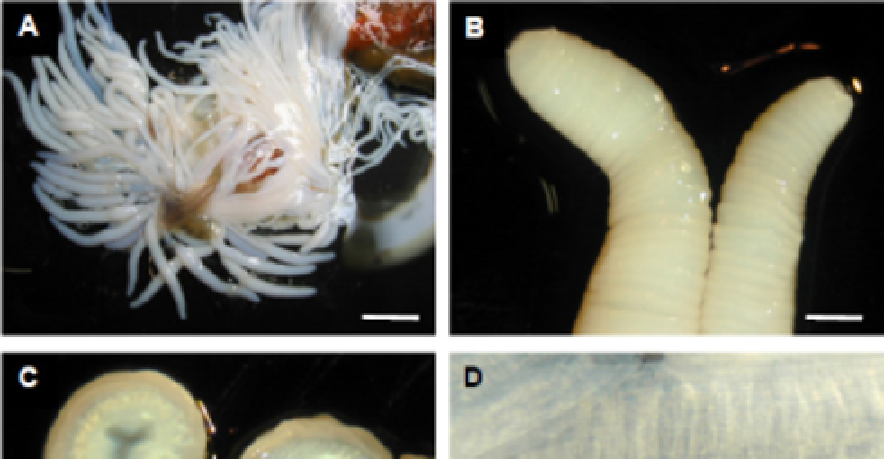
3.1 Tubule structure and microscopy
Dissection of euthanised
H. dofleinii
showed that the body cavity contained a large number,
several dozen, Cuvierian tubules in their compressed form (Figure 1A). In their compressed
form the tubules were not sticky, but they rapidly became sticky on mechanical extension,
even from the dead animals. The compressed, individual tubules showed a corrugated and
folded surface (Figure 1B), which would allow extension when required, like a concertina
bellows. In cross-section (Figure 1C) a three-lobed channel could be seen that would allow
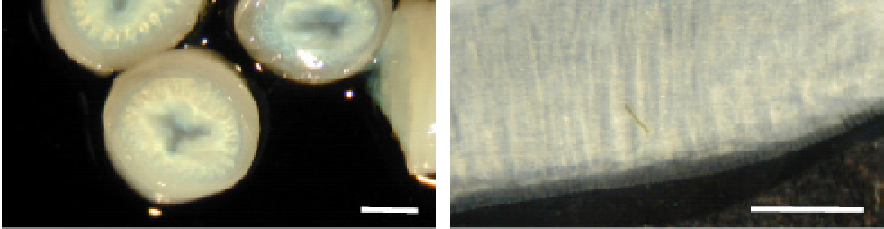
fluid insertion for expansion of the tubules. When expelled and fully extended
in vivo
, these
tubules became instantly sticky and changed from the 25-35 mm compressed length up to
around 350-400 mm. The fully extended tubules (Figure 1D) were typically about 4mm flat
width when fully inflated, and still showed some patterning from the folding that was
present in the compressed state. Individual animals contained several dozen non-inflated
tubules, but when the animals were stimulated only a small number were expelled,
normally around 8-12.
Fig. 1. Cuvierian tubules for
H. dofleinii
. (A, B, C) After dissection of a euthanised animal,
showing, (A) the total mass of tubules, (B) the tips of compressed tubules, and (C) the cross-
section of compressed tubules. (D) The surface of an
in vivo
expelled tubule. Bar (A) = 10
mm, Bars (B, C, D) = 1 mm.