Biomedical Engineering Reference
In-Depth Information
The MTT tests were widely used cytotoxicity tests because they are easy, fast and cheap. But,
in the case of Mg materials, the use of these test kits leads to false positive or false negative
results. It is conceivable that Mg in the highly alkaline environment may be able to open the
ring form of the tetrazolium salt and bind to it, which could lead to a change in colors similar
to the formation of formazan in the case of the MTT tests with cells [20]. Thus the results of
cytotoxicity tests conducted by MTT test will be higher than the true story. It is well known
that the neutral red assay and the MTT assay exhibited almost identical reaction patterns for
most test materials in L929 cells [33]. Thus, in this study, the neutral red kits were used to
assay the cytotoxicity. Its measurement principle is based on the uptake of the vital dye neutral
red into lysosomes of viable cells. Neutral red is accumulated because of the low intravesicular
pH value. Lysosomes are, however, only one type of subcellular compartments which are
acidified by ATP-driven proton pumps (V-type ATPase) and related low intravesicular pH
value across vesicle membranes [34]. Accumulation of neutral red in acidic intracellular
vesicles needs both ATP as a universal metabolic energy source for proton translocation
against an electrochemical H
+
-gradient and tightly sealed vesicle membrane to maintain
potential differences. In an alkaline environment with Mg
2+
, neutral red could lead to a change
in color to yellow. But, the uptake of the vital dye neutral red into lysosomes was red color.
The preliminary results of this test show that it seems not to be influenced by corroding Mg.
Therefore, the neutral red assay may be regarded as a valid alternative method to determine
cell viability, as it shows no interference with the corroding materials. The in-vitro cytotoxicity
of Mg-4.0Zn-0.2Ca alloy was found to be Grade 0-1, indicating that the alloy was bio-safe.
3.5 In-vivo degradation
Furthermore, in order to further study the biocompatibility of Mg-4.0Zn-0.2Ca alloy, the in-
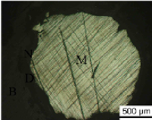
vivo test was conducted on this new type magnesium alloy. Fig.15 showed the optical images
of the cross-section of bone and magnesium implants after 3 months implantation. It could be
seen that all the shapes of the magnesium implant had been changed from rod shape to
irregular shape, indicating the implant was corroded by the body fluid, or the implant
degraded in the body fluid. Meanwhile, a degradation layer or a reaction layer could be clearly
found on the surface of the alloy implant, as indicated by D in Fig.15. In addition, newly
formed bone was observed between the degradation layer and bone tissue around the
magnesium alloy implants, as shown by N in Fig.15. The degradation rate was calculated
according to the ratios of the cross section area of the residual implant to the original implant.
After 3 months implantation, about 35-38% Mg-4.0Zn-0.2Ca alloy implant was degraded.
Significant difference (p < 0.05) in the in-vivo degradation rates was observed.
Fig. 15. Optical images of the cross-sections of Mg-Zn-Ca implants and bones after 3 months
post implantation (M, metal; D, degradation layer; N, new bone; B,bone).