Biology Reference
In-Depth Information
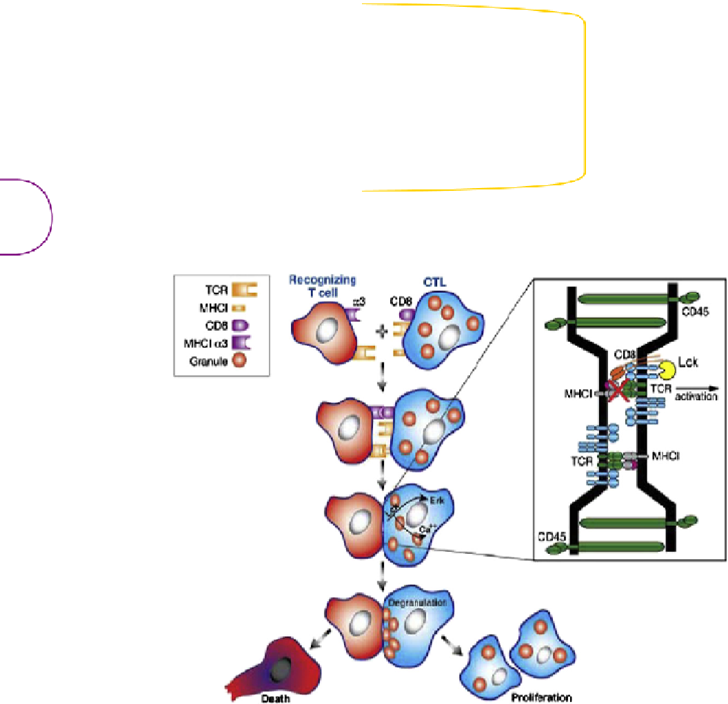
a proliferative response in the veto effector cells, granule polarization at
the interface of the veto cell with the cytotoxic effector cell, and release
of perforin by the veto cell, which then induces apoptosis in the cytotoxic
effector cell (
Figure 5.3
).
Effector
Veto
MHC
class I
FIGURE 5.2
Signaling in recipient anti-donor effector T cells
induced by donor veto cells. Binding of CD8
molecules on the donor veto cell with the
α
3 domain
of MHC class I molecules on the recipient effector cell,
together with T-cell receptor signaling in the recipi-
ent effector cell, induces Mek-Erk activation, which
decreases XIAP levels and their inhibitory effect on
caspase activation, thereby enabling FasL-mediated
apoptosis (adapted from Reich-Zeliger et al.
[129]
,
with permission).
α3
CD8
Mek-Erk
MHC
class I
TCR
XIAP
DISC
Fas
FasL
Caspases
Cell Death
108
FIGURE 5.3
Signaling in donor veto cells recognized by recipient anti-donor effector T cells. Binding of the
α
3 domain of MHC class I molecules on the recipient effector cell
(red) with CD8 molecules on the donor veto cytotoxic T cell (blue) induces Lck activation and Ca
2+
release, followed by granule polarization and degranulation at the
interface with the effector cell. The T-cell receptor of the donor veto cytotoxic T cell does not recognize the recipient effector cell (inset). Nonetheless, trapping of T-cell
receptors in lipid rafts formed by aggregation of CD8 molecules could theoretically lead to Src kinase activation and Erk phosphorylation, thereby providing signals that
promote survival and proliferation of the veto cell (adapted from Milstein et al.
[130]
, with permission).


Search WWH ::

Custom Search