Biology Reference
In-Depth Information
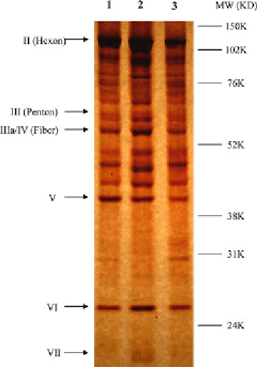
Fig. 2
Silver staining of Adenoviral capsid proteins. Composition of Ad5 and
hexon modifi ed adenoviral vectors analyzed by silver stain. 5 × 10
10
denatured
viral particles were loaded per lane and run on a 9 % SDS-polyacrylamide gel,
and stained using Pierce silver staining kit following manufacturer instructions.
Lane (1) Ad5-CMVLacZ, Lane (2) Ad5-CMV-LacZ with point mutations in HVR5,
Lane (3) Ad5-CMV-LacZ with point mutations in HVR7
We have recently been using the NanoSight™ platform as a tool for
assessing viral quality and titers, and fi nd that it accurately refl ects
total particle count as gauged by viral microBCA protein assay. It
also provides additional information on particle monodispersity
and potential aggregation, and we have also found it to be a useful
tool for studying virus: host protein interactions. The following
gives an overview of how we use the machine, however, we recom-
mend full, hands-on training from a NanoSight™ expert, as well as
careful reading of the provided manual prior to commencing
experiments. Example screen shots and data are shown in Fig.
3
.
3.2.2 NanoSight™ for
Assessing Viral QC
1. With the computer switched on, open NTA software.
2. Dilute virus prep 1/1,000 in PBS, so the concentration is
within the defi ned detection range (10
6
to 10
10
particles per
mL—occasionally further dilution will be necessary). A mini-
mum total volume of ~1,000
μ
L is required (hence 1
μ
L of
viral stock solution) (
see
Note 6
).
3. Undo screws holding the lid in place, clean both glass plates
with ethanol and lint free paper, then replace the lid and screw
back into place.