Biology Reference
In-Depth Information
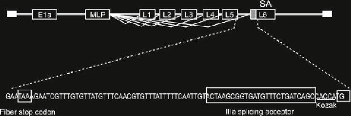
Fig. 1
IIIa splicing acceptor gene expression system. A transgene is expressed as a new late transcriptional
unit regulated by the MLP. The splicing acceptor from the adenovirus gene IIIa is located right after the fi ber
polyA
adenovirus death protein) is an option to create space for exoge-
nous transgenes. However, this E3 deletion may accelerate virus
clearance in an immunocompetent host [
3
]. As splicing acceptors
are much shorter (26bp) compared to IRES (500 bp), they repre-
sent our preferred option to arm adenoviruses with transgenes.
Another option that also saves cloning space for transgenes is to
link the transgene to a virus protein using the 2A sequence that
during translation precludes the formation of the peptide link [
4
]
and produces two separate proteins from the same mRNA.
We have used the adenoviral splicing acceptor IIIa after the
fi ber open reading frame to express transgenes. This acceptor cre-
ates a new splicing unit in the long major late transcript (which
would correspond to L6), without affecting the virus cycle [
5
,
6
].
We also usually insert the Kozak sequence (CCACC) before the
ATG of the start codon of the gene of interest to improve transla-
tion initiation. Finally, we use the TAA stop codon to terminate the
transgene and add TAAA after it to form a polyA signal (
see
Fig.
1
).
Late gene expression is an advantage when the proteins encoded
by the gene of interest can interfere with the viral cycle, such as
suicide, proapoptotic, or cytotoxic proteins.
In this chapter we describe basic protocols for the characteriza-
tion of oncolytic adenoviruses. We have not included virus con-
struction and purifi cation protocols as those are described in other
chapters of this topic. The infectious unit titer and the IC50 assays
are used to measure the concentration of active virus in a sample.
A differential infectious unit titer in two cell lines can be used to
assess the relative permissiveness of a given virus in these two cell
types. If a given cell line is infected with several viruses at the same
multiplicity of infection (MOI or infectious units/cell) or with the
same physical particle titer (vp/cell), the IC50 refl ects the propa-
gation effi cacy (in vitro oncolytic potency). The size of a plaque
may also indicates this propagation effi ciency, but it is less quantita-
tive and more dependent on the cell line used, because cells that do
not detach easily do not form clear plaques. In vivo, the follow up