Biology Reference
In-Depth Information
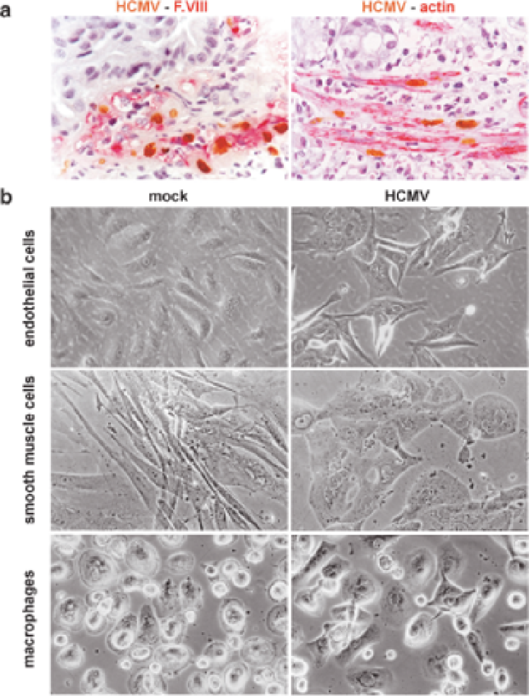
Fig. 1
a
Immunohistochemical evidence of productive infection in endothelial cells and smooth
muscle cells in vivo, as indicated by focus formation within the respective cell layers.
Brown
nuclear signals
, detection of HCMV immediate early antigens by indirect immunoperoxidase
labeling;
red cytoplasmic signals
, detection of F.VIII-related antigen (endothelial cells) and actin
(smooth muscle cells) by indirect immunoalkaline phosphatase labeling;
blue nuclear signals
,
counterstaining with hematoxilin.
b
Phase contrast micrographs of HCMV-infected cell cultures.
Irrespective of the great morphological differences between cultured cells prior to infection,
HCMV productive replication results in uniform morphological appearance with cytomegaly and
nuclear inclusions