Biology Reference
In-Depth Information
b
In Utero
Decidua
Invasive CTBs
CC
Decidual
cells
a
CMV ICP
/CK/
TO-PRO
CTB
c
d
60 min p.i.
60 min p.i.
NK
BV
VC
AV
ST
2
Infected
invasive
CTBs
FV
Tol
rec
/EGFR/
To-PRO
Tol
rec
/
α
1
β
1/
TO-PRO
Differentiated
CTBs
e
f
60 min p.i.
24 h p.i.
In Vitro
Tol
rec
/
α
V/
TO-PRO
CMV IE1&2
/CK
Fig. 4
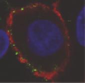
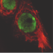
CMV replicates in invasive cytotrophoblasts that express virion receptors.
a
Diagram of
differentiating/invading cytotrophoblasts. The
blue field
represents the localization of viral pro-
teins in
b, c, d, e,
and
f
.
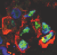
b
Invasive cells in decidua infected in utero. The inset corresponds to the
outlined area in the panel. Toledo
rec
virions (
green
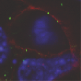
) bind to cytotrophoblast membranes with EGFR
(
c
), integrin α1β1 (
d
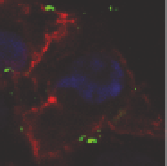
), and integrin αV (
e
) (
red
) at 1 h after infection.
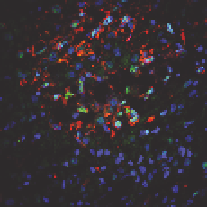
f
IE1 and IE2 proteins
(
green
) in cytokeratin (
CK
)-stained cells (
red
) at 24 h after infection. Nuclei were counterstained
with TO-PRO-3 iodide (
blue
)
the invasive pathway, integrins α1β1 and αVβ3 are upregulated (Damsky et al. 1994;
Zhou et al. 1997). Location of CMV-infected invasive cytotrophoblasts is illustrated
in Fig. 4a. Analysis of early gestation decidua from a placenta with low neutralizing
titers showed that infected cytotrophoblasts express viral replication proteins
(Fig. 4b). Cultures of differentiated cytotrophoblasts on filters coated with Matrigel
were used to monitor these cells for expression of potential receptors, binding of
Toledo
rec
virions and productive infection. Virions (punctate green) adsorbed to
membranes of cells expressing EGFR (Fig. 4c) and integrins α1β1 (Fig. 4d) and αV
(Fig. 4e) at 1 h after infection. In addition, CMV IE1- and IE2-infected cell proteins
were detected in nuclei of Toledo-infected cytotrophoblasts at 24 h, suggesting
early-stage viral replication (Fig. 4f). These results showed that virion attachment
and infection of invasive cytotrophoblasts occur in vitro in cells that express CMV
receptors, EGFR and integrins αV and α1β1.
Using function-perturbing antibodies and soluble integrins, we evaluated the role
of molecules that function as potential receptors in differentiating/invasive cytotro-
phoblasts (Maidji et al. 2007). Intense expression of EGFR and integrins α1β1 and