Biology Reference
In-Depth Information
b
EGFR
-
Villus
CTB
In Utero
VC
Cell column
CTB
CC
CC
a
Proximal
Distal
CTB
BV
VC
EGFR
/gB/
TO-PRO
Cell column
CTB
c
ST
AV
FV
VC
CC
Infected
invasive
CTB
α1β1
+
CMV gB
/
α
1
β
1/
TO-PRO
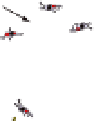
Fig. 3
CMV virions bind to cytotrophoblast outgrowths in a model of anchoring villus develop-
ment in vitro.
a
Diagram of an anchoring villus cell column. The
blue field
represents the localiza-
tion of virions in panels
b
and
c
. The insets in panels
b
and
c
correspond to the outlined areas in
each panel. Explants were fixed at 24 h postinfection.
b
Cell column cytotrophoblasts did not react
with antibodies to EGFR (
red
).
c
Toledo
rec
virions (
green
) bind cytotrophoblasts expressing α1β1
integrin (
red
) on the edge of a distal cell column. Nuclei were counterstained with TO-PRO-3
iodide (
blue
)
cytotrophoblasts in these outgrowths did not express EGFR (red) (Fig. 3b).
Nonetheless, virion attachment was observed as punctate (green) staining in membranes
of distal columns where potential integrin receptors αV and α1β1 were expressed.
Cytotrophoblasts on the edge of distal portions of columns upregulated α1β1 (red),
and Toledo
rec
virions were bound to surface membranes (Fig. 3c). Integrin αV was
also present (data not shown). The results suggest that cytotrophoblast expression
of integrin coreceptors without EGFR enables virion binding but is insufficient for
entry and replication.
Replication in Differentiating/Invading Cytotrophoblasts
Analysis of first- and second-trimester placentas from women with moderate CMV
neutralizing titers showed that interstitial cytotrophoblasts in decidua are infected
in utero (Pereira et al. 2003). As cytotrophoblasts differentiate and progress along