Biology Reference
In-Depth Information
CC
a
CTB
BV
VC
In Vitro
ST
AV
FV
b
c
ST
ST
CTB
CTB
Tol
rec
/
α
V/
TO-PRO
Tol
rec
/EGFR/
TO-PRO
d
ST
Tol
rec
CTB
α
V
EGFR
Fig. 2
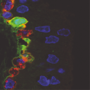
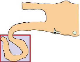
CMV replicates in cytotrophoblasts expressing EGFR and αV integrin in villous explants
infected in vitro.
a
Diagram shows focal infection in a floating villus. The
blue field
represents the
localization of CMV structural and replication proteins in panels
b
and
c
. Explants were fixed at
24 h postinfection.
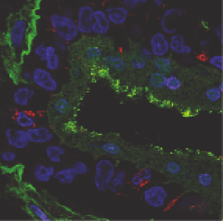
b
Toledo
rec
virions bound to apical microvilli of syncytiotrophoblasts (
ST
).
Selected cytotrophoblasts (
CTB
), but not syncytiotrophoblasts, reacted with antibody to αV
integrin (
red
).
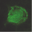
c
Underlying cytotrophoblasts stained intensely with EGFR-specific antibody (
red
);
replicating Toledo
rec
broadly expressed green fluorescent protein in nuclei of infected cells. Nuclei
were counterstained with TO-PRO-3 iodide (
blue
).
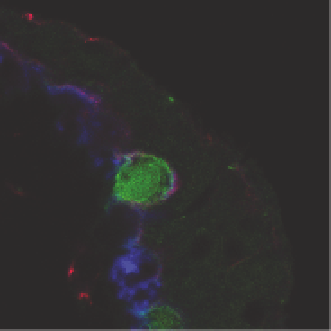
d
Villous cytotrophoblasts infected with
Toledo
rec
simultaneously expressed αV integrin (
red
) and EGFR (
blue
)