Biology Reference
In-Depth Information
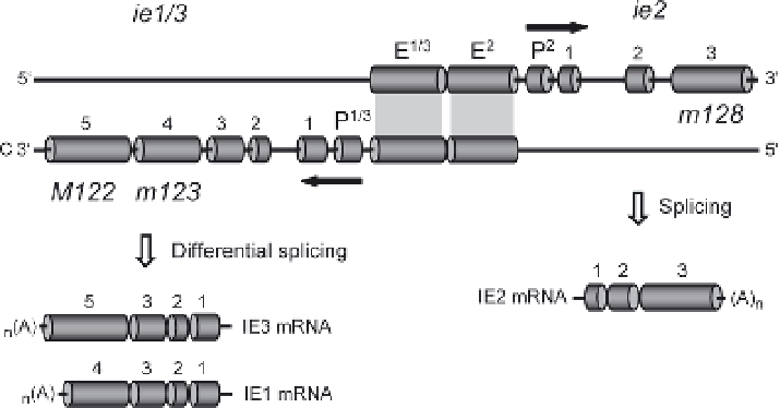
Fig. 1
Bidirectional gene pair architecture of the mCMV MIE locus.
Arrows
indicate the direction
of transcription. Numbered cylindrical boxes represent exons.
C
, complementary strand.
M
and
m
indicate mCMV ORFs homologous to hCMV ORFs and mCMV private ORFs, respectively,
according to the nomenclature used by Rawlinson et al. (1996).
P
promoter,
E
enhancer
The 76/89-kDa IE1 protein is involved in breaking epigenetic host cell defense
by early disruption of nuclear domains (ND)10 (see the chapter by G. Maul, this
volume); it co-transactivates the expression of viral early (E)-phase genes and
autostimulates its own promoter. Interestingly, it also acts as a transactivator of
cellular genes involved in dNTP biosynthesis, such as thymidylate synthase
(Gribaudo et al. 2000) and ribonucleotide reductase (Lembo et al. 2000), a prop-
erty thought to facilitate virus replication in resting cells (for reviews, see
Simon et al. 2006b; Tang and Maul 2006). Clearly, efficient provision of dNTPs
could possibly be a key parameter in virus reactivation from latently infected
cells, which are most likely resting cells, and it will be intriguing to test this
idea. The importance of the 88- to 90-kDa IE3 protein for virus reactivation is
undoubted, as IE3 is the essential transactivator of viral E gene expression
(Angulo et al. 2000). Thus, beyond MIE locus transcription initiation, differen-
tial splicing generating IE3 mRNA is a crucial second molecular checkpoint
in the transition from mCMV latency to reactivation. Strikingly, no essential
function could so far be identified for the 43-kDa IE2 protein (Cardin et al.
1995; Messerle et al. 1991). It remains an open question whether IE2 is really
dispensable or whether we just failed to ask the correct questions, to design the
proper experiments, and to look at the right place and time. In this context, it is
of interest that recent work by Ishiwata et al. (2006) has localized IE2 protein
in the brain of neonatally infected mice at a stage of prolonged infection selec-
tively in neurons of the cortex and hippocampus, while the IE3 protein was
preferentially expressed in glial cells only at an early stage of the infection. It is
a challenge to identify the function of IE2 and to understand why its expression