Biology Reference
In-Depth Information
1985; Gilbert et al. 1989), which strongly suggested that viral transmission was
cell-based and not mediated by free virus. Consequently, one of the cellular sites of
latency was believed to be in the peripheral blood compartment.
We now know that the latent load of HCMV in healthy carriers is around 1
genome-positive cell per 10,000 peripheral blood mononuclear cells (PBMCs)
(Slobedman and Mocarski 1999), clearly below the detection limits of Northern,
Southern and Western analyses. However, the use of a highly-sensitive PCR
approach finally permitted the analysis of HCMV latency in vivo and defined the
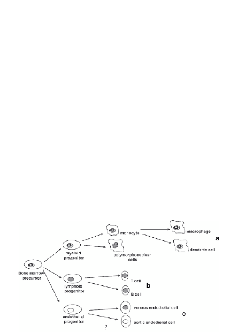
myeloid lineage as an important site of HCMV latency (Fig. 1). An experimental
approach that isolated different fractions of cells from the blood of healthy seroposi-
tives showed that carriage of HCMV DNA occurred predominantly in the leukocyte
fraction of peripheral blood - particularly in the CD14
+
monocyte population (Fig.
1a). HCMV was not found in the lymphocyte fraction or the polymorphonuclear
cells (Fig. 1b) (Bevan et al. 1991; Taylor-Wiedeman et al. 1991, 1993; Stanier et al.
1992). Monocytes, however, represent a short-lived continually renewable population
of cells that arise from haematopoietic cell precursors (CD34
+
cells) present in the
bone marrow (Metcalf 1989). These cells were also shown to be HCMV genome-
positive, suggesting that the bone marrow represents one latent reservoir of virus
(Mendelson et al. 1996; Sindre et al. 1996). Although CD34
+
cells are sites of
latency for HCMV and are a common precursor of both lymphoid and myeloid cells,
the carriage of virus appears to be restricted to cells of the monocyte/myeloid lineage
by, as yet, undefined mechanisms (for review see Sinclair and Sissons 2006).
Fig. 1
HCMV latency is established in bone marrow progenitors and is carried in the myeloid
lineage. During natural latency, HCMV DNA can be detected in bone marrow progenitor cells that
give rise multiple lineages. However, the carriage of viral genomes is detected in the myeloid line-
age (
a
) and not the lymphoid lineage (
b
). A third endothelial lineage (
c
) has been proposed, but
not proven (
?
), which may also provide another site of HCMV latency in aortic, and not venous,
endothelial cells