Biology Reference
In-Depth Information
a less structured tegument layer, and bounded by a trilaminate membrane envelope
(Fig. 1a). The HCMV genome is composed of a linear, double-stranded DNA mole-
cule (236 kbp in wild type virus), the largest among the human herpesviruses, and
over 50% larger than that of herpes simplex virus type 1 (HSV-1) (see the chapters by
E. Murphy and T. Shenk, and G.S. Pari, this volume). The capsid is isosahedral and
about the same diameter as that of HSV (~110 nm, depending on preparation).
Accommodating a larger DNA in a similar diameter capsid may be achieved by
eliminating the maturational protease (pUL80a) from the interior of CMV capsids
(Chan et al. 2002; Loveland et al. 2007). The capsid is composed of four integral
protein species (for HCMV, pUL46, pUL80.5, pUL85, pUL104) that are organized
into 162 capsomeres (150 hexamers plus 12 pentamers) and 320 triplexes located
between the capsomeres. By analogy with HSV-1, one of the pentamer positions is
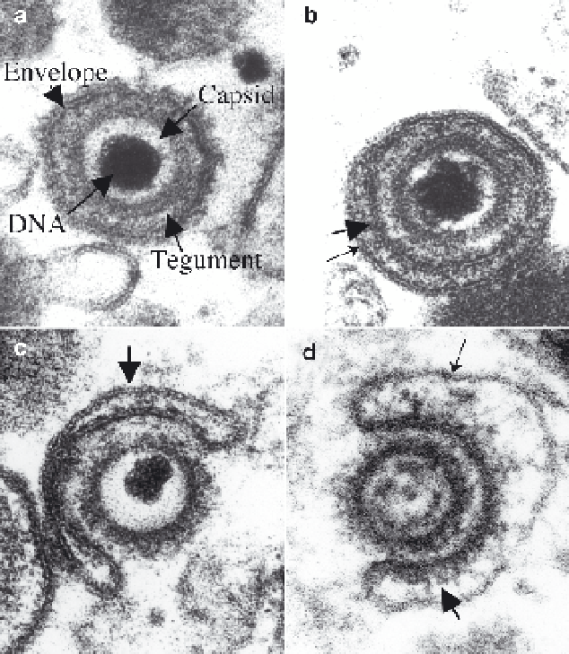
Fig. 1
Particles in cytoplasm of CMV-infected cells. Shown here are electron micrographs of
a
virion with DNA, capsid, tegument, and envelope indicated by arrows,
b
virion within small
vesicle or tubule indicated by thinner arrow,
c
tegumented C-capsid, with coarse fibrillar material
especially evident on right-hand side, budding into a vesicle or tubule (
arrow
) to become virion,
and
d
tegumented B-capsid budding into large tubule or vesicle (
top arrow
) to become NIEP;
showing thickening of vesicle membrane where apposed to particle (
bottom arrow
)