Biology Reference
In-Depth Information
3
10
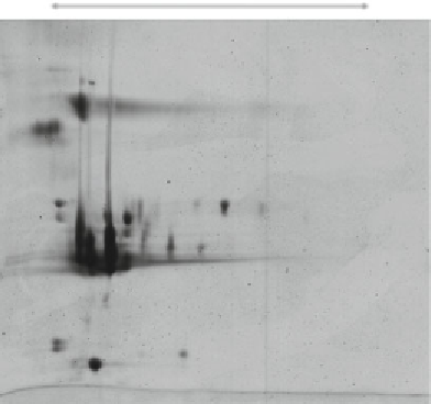
Fig. 1
2D gel electrophoresis of champagne base wine proteins stained with
colloidal Coomassie Brilliant Blue. Wine proteins were separated by IEF in 7 cm
long pH 3-10 IPG strips, followed by SDS-PAGE in vertical 12 % gels. Champagne
base wine proteins are mainly located between pH 3 and 5
3.5 Second
Dimension (SDS-PAGE)
1. Prepare the 12 % polyacrylamide gels (composition of two
gels, 1 mm width, for Mini-Protean 3 system, Bio-Rad) by
mixing slowly 4.875 mL of deionized water, 5.4 mL of 1 M
Tris-HCl, pH 8.8 buffer, 4.5 mL Acrylamide-Bisacrylamide
40 % ratio 37.5:1 (Bio-Rad), 150
μ
L of SDS 10 % (w/v) in a
25 mL glass beaker. Add 75
L
of TEMED. Cast gel in a 7.25 cm × 10 cm × 1 mm gel cassette.
Allow space (
μ
L of APS 10 % (w/v) and 7.5
μ
1 cm) for the IPG strip and cover the gel with a
few milliliters of water-saturated
n
-butanol or deionized water.
2. After polymerization (
≈
≈
1 h at room temperature), discard
water-saturated
n
-butanol and wash thoroughly with deion-
ized water.
3. Pipet the overlay agarose solution, previously warmed, on the
top of the vertical gel and place immediately the IPG strip in
close contact with the SDS polyacrylamide gel (
see
Note 15
).
4. Let the overlay agarose solution to set at room temperature for
at least 1 min.
5. Run the electrophoresis at 30 V during 20 min, then at 175 V
until the front dye reached 5 mm from the bottom of the gel.
6. The gels were stained with colloidal Coomassie Brilliant Blue
according to Candiano et al. [
29
]. We were able to visualize
around 30 and 50 spots on pH 3-10 (Fig.
1
) and pH 3-6
(Fig.
2
) gradients, respectively.