Biology Reference
In-Depth Information
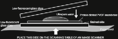
Fig. 1
Detection of fl uorescence of Cy5 and ECL-double stained membrane. After
incubation with ECL plus (plex) substrate, place the membrane on a low-
fl uorescent glass plate with the stained side on which the proteins are stuck
facing down. Drip a few milliliters of water on the back of the membrane and
place another glass plate on the membrane to allow the water to spread over the
entire surface of the membrane to remove air bubbles
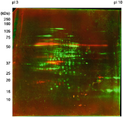
Fig. 2
A merge image of all soluble rice seed protein- and IgE-binding protein-
patterns. All transferred rice seed proteins were labeled with Cy5, and the IgE-
binding proteins were labeled with ECL after incubation with the serum of a
patient with rice allergy (diluted 1:100) followed by that with HRP-linked anti
human IgE (diluted 1:1,000). The merged scan image of the Cy5 (
green
) and ECL
(
red
) images is shown
4. Dehydrate the gel pieces with dehydration buffer followed by
acetonitrile, and air-dry.
5. Add 10
L of Trypsin
Gold in 0.01 % Protease Max solution) to the completely dried
gels. After keeping the tubes on ice for 15 min, incubate them
for 2 h at 37 °C.
μ
L of trypsin digest solution (20 ng/
μ
3.5 Mass
Spectrometry and
Protein Identifi cation
1. Desalt the tryptic digests with Zip-Tip C18.
2. Mix 0.5
μ
L of digest and 1
μ
L of
α
-CHCA and spot onto an
Opti-TOF 384 well Insert plate.