Biology Reference
In-Depth Information
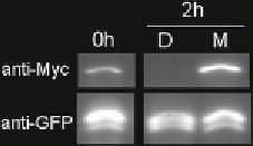
Fig.
3
Inhibition of proteasome-dependent protein degradation with MG132
treatment. The crude extract (0 h) from tobacco leaf was incubated with DMSO
(D) or MG132 (M) for 2 h and used for western blotting analysis with anti-Myc
and anti-GFP antibody. Overexpression of ubiquitin ligase ATL31 promoted
proteasome-dependent degradation of the specifi c target protein Myc-14-3-3
with DMSO treatment for 2 h, whereas MG132 treatment inhibited the degrada-
tion of the target. GFP was used as a control for normalization of amount of the
expressed proteins
χ
5. After blocking treatment by incubation with blocking buffer,
incubate the membrane with 1/5,000-diluted anti-Myc or
anti-GFP antibody with PBS-T for 1 h in room temperature
with shaking.
6. Remove the antibody and wash the membrane with PBS-T
buffer.
7. Incubate the membrane with 1/25,000 HRP-labeled anti-
mouse IgG antibody for 1 h in room temperature with
shaking.
8. Remove the antibody and wash with PBS-T buffer.
9. Add the detection solution onto the membrane and incubate
for 5 min.
10. Detect and quantify the signal with luminescent image
analyzer LAS3000 (Fujifi lm, Tokyo, Japan) (
see
Fig.
3
and
Note 9
).
3.2 In Vivo MG132
Treatment and
Detection of
Ubiquitinated Protein
1. Sterilize
Arabidopsis thaliana
seeds and sow seeds on petri dish
with MS medium.
2. Germinate the seedlings and let grow for 10 days (
see
Note 10
).
3.2.1 Preparing Plants
1. Add MG132 to 1/2 MS liquid medium to a fi nal concentra-
tion of 50
3.2.2 MG132 Treatment
for Plants
M. Also, add DMSO of the same volume of the
MG132 to 1/2 MS liquid medium as a negative control.
2. Pour 3 mL, respectively, of 1/2 MS liquid medium containing
MG132 or DMSO into a 35 mm petri dish.
3. Float three plants on 1/2 MS liquid medium in each dish.
Incubate dishes under normal growth conditions for 15 h (Fig.
4
).
4. Wipe attached medium from plants and weigh them.
μ