Biology Reference
In-Depth Information
×
10
4
8
Gl NAc
c
Mannose
Xylose
6
Fucose
Galactose
4
2
0
1400
1600
1800
2000
2200
2400
2600
m/z
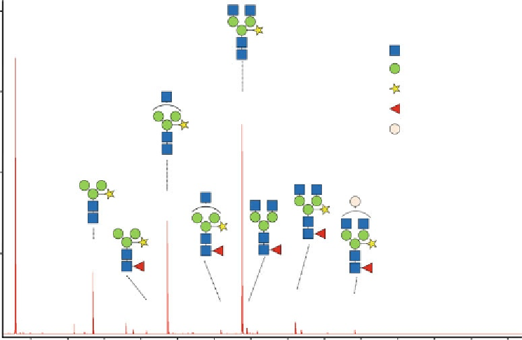
Fig.
2
A typical mass spectrum of
N
-glycans of AmyI-1, obtained by the glycoblotting-mass spectrometry
method. All molecular weights represent monoisotopic masses of the respective [M+aoWR-H
2
O+H]
+
ions of
glycan species. aoWR,
m/z
447.22; H
2
O,
m/z
18.01. Structural annotation of peaks detected in the MALDI-TOF
MS were obtained using the GlycoMod tool online database
3.6
N -Glycome in
Major
N
-glycoforms of rice secretory AmyI-1 has been determined
using traditional aminopyridine (PA)-derivatization and multidimen-
sional HPLC analysis [
3
]. The same AmyI-1 glycoprotein sample was
subjected to the glycoblotting-mass spectrometry technique, and the
obtained results are shown in Fig.
2
and Table
1
. Several fucose-con-
taining glycans (i.e., Man
3
GlcNAc
4
Fuc
1
Xyl
1
, Man
3
GlcNAc
3
Fuc
1
Xyl
1
,
Man
4
GlcNAc
4
Fuc
1
Xyl
1
, Man
3
GlcNAc
2
Fuc
1
Xyl
1
, Man
3
GlcNAc
3
Fuc
1
)
were newly detected using the glycoblotting method.
Rice
-Amylase I-1
(AmyI-1)
α
4
Notes
1. The “glycoblotting” technique is applicable to a wide range of
protein amounts (10
−6
-10
−3
g). Use of detergent is allowed,
but its concentration must be as low as possible (e.g., Triton
X-100 should be less than 0.5 M).
2. The step involving the reduction and alkylation of proteins may
be skipped when the protein sample is dissolved in solution.
3. The common
N
-glycan releasing enzyme, PNGase F, should not
be used, because this enzyme cannot digest the plant
N
-glycans
with conjugation of
α
1,3-fucose to the core structure.