Biology Reference
In-Depth Information
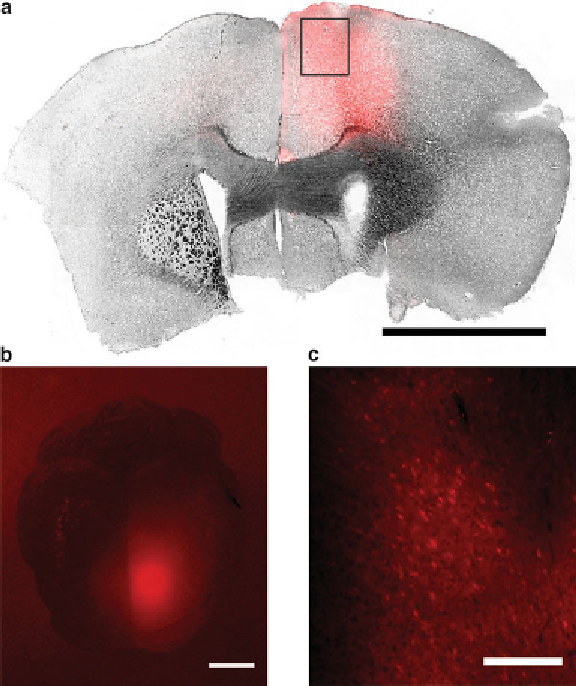
Fig. 3
Expression pattern following 1
- ChR2(H134R)-
mCherry into the mouse motor cortex. Virally transduced cells exhibit
red
fl uores-
cence due to mCherry expression. (
a
) Coronal brain section showing injection
center (identifi ed by injection tract) approximately 1 mm anterior to bregma. Brain
injury by the optic fi ber is visible at the upper edge. Scale bar: 2 mm. (
b
) Dorsal
view of the brain prior to sectioning. Scale bar: 2 mm. (
c
) Close up of cells [
box
in (
a
)]. Somata of transduced cells are clearly visible. Scale bar: 200
μ
l injection of AAV5-CaMKII
α
μ
m
4. Anesthesia induction can be performed by placing the mouse
in an induction box with 4 % isofl urane. However, light ket-
amine/xylazine anesthesia will allow for easier handling until
the mouse is placed on the stereotactic device and deeply anes-
thetized by isofl urane application.
5. The medial-lateral position normally does not require any
readjustment if the ear bars were positioned evenly. Use the
ear bar scales to verify even positioning.
6. It is advisable to fi rst align height of lambda and bregma visu-
ally by looking at the skull from the side. If this is done cor-
rectly, little adjustment will be needed afterwards.
Search WWH ::

Custom Search