Biology Reference
In-Depth Information
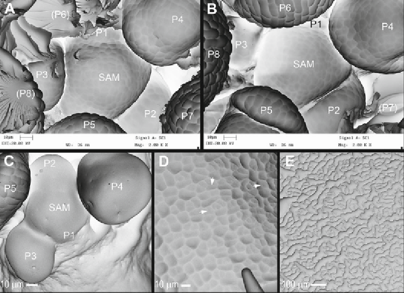
Fig. 3
SEM micrographs of casts representing apices of infl orescence shoots (
a
-
c
) or leaf epidermis (
d
,
e
) of
A. thaliana
. (
a
,
b
) Two casts obtained from the same, high-quality mold, which were trimmed in a different way.
Flower primordia are labelled in both casts with the same symbols (
P
and the number increasing with the
primordium age). (
a
) Cast trimming enables better visualization of the shoot apical meristem (SAM): the cell
outlines are more apparent and more of the SAM surface can be examined than in the other cast obtained from
this mold, because all the older primordia were cut off with razor blade (their symbols are in
parentheses
).
However, the surface of the SAM is locally damaged between primordia P1 and P3. (
b
) The specimen is
charged because of a deep grove between primordia and the SAM. Parts of the SAM periphery, as well as the
primordium P1, are hidden behind surrounding primordia. (
c
) The cast obtained from a mold of insuffi cient
quality, in which the dental polymer has not set properly, presumably because of earlier plant reaction to
aphids. Before the mold was taken, some older primordia were removed (
lower right
part of the image), and
the released cell sap probably affected the polymer setting. Unlike this apex portion, the surface of P5 is rather
well represented. (
d
,
e
) Casts from molds taken from the adaxial epidermis of the same leaf at 6 days time
interval. In the younger epidermis (
d
) cell outlines are less prominent than in older (
e
). Moreover, in the younger
epidermis (
d
), cells are still dividing and the younger anticlinal walls (e.g., those pointed by
arrows
) are much
less distinct than older walls, unlike the walls in older nearly differentiated epidermis (
e
). All the images were
taken with the aid of SEM machine LEO435VP
a stereopair of images has to be taken [
7
,
8
]. These are two
images of the same region taken at different SEM stage tilt
angles (we use a 10° difference in tilt for shoot apices or leaves).
After taking one image, tilt the stage precisely with respect to
the
X
-axis (
see
Note 12
) and then move the specimen along
the
Y
-axis to come back to the region of interest. During this
Search WWH ::

Custom Search